Membrane Structure and Dynamics
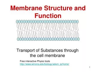
Membrane Structure and Dynamics. Membrane functions - physical barrier from entry and exit form cell and organelles What are membranes - Lipid bilayers with proteins imbedded or associated on either side of the membrane

Membrane Structure and Dynamics
E N D
Presentation Transcript
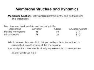
Membrane Structure and Dynamics Membrane functions - physical barrier from entry and exit form cell and organelles What are membranes - Lipid bilayers with proteins imbedded or associated on either side of the membrane Ions and polar molecules basically impermeable to membrane - energy costs too high
Membrane components - • 60 to 70% of mammalian lipids are phospholipids • Bacteria have almost no PC and are mostly PE • Neuronal tissue (myelin) PI > PC • Alterations in lipid composition - permeability, fluidity, exocytosis, neural transmission and signaling potential
Membrane Asymmetry • P-ethanolamine and P-serine predominately faces inside of cell • P-choline faces outside of membrane and inside of organelles • carbohydrates of glycoproteins facing outside • During apoptosis there is a re-arraignment of lipids where phosphatidyl serine moves to the exterior face of the membrane. One of the key signals of cell death
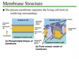
Membrane Fluidity - Singer and Nickolson fluid mosaic model - allows for dynamic nature of membrane - little transition of lipids can take place without specific enzymes to mediate transfer - flipase
Proteins - Add function and structure to membrane • Extrinsic proteins (peripheral) • Loosely attached to membrane • ionic bonds with polar head groups and carbohydrates • hydrophobic bonds with lipid • proteins have lipids tails • easily displaced from membrane • salt, pH, sonication
Transmembrane portion often a helix • takes about 20 aa to cross membrane • many proteins cross many times • odd # of transmembrane regions, why • -COOH terminal usually cytosolic • -NH3+ terminal extracellular • can be predicted by amino acid sequence • high % of side chains will be hydrophobic • Hydropathy scale used to predict • free energy change - from organic to water • long regions unusual in soluble proteins • Non membrane sections often modified • lipid, carbohydrate
Intrinsic proteins - tightly bound to membrane - span both sides Protein has both polar and hydrophobic sections removed only through disrupting membrane with detergents detergents disrupt lipid bilayer and incorporate proteins and some lipids into detergent micelles • allows for purification of membrane proteins • reconstitute into specific vesicles for study
Membrane associated proteins N or C terminal modifications Tightly associates protein to membrane Isoprenylated at C Terminus -Geranylgeranyl and farnesyl groups - from cholesterol biosynthesis - Lovastatin inhibits post-translational modification - deterimined for Ras and pancreatic cancer. -CAAX box - C = Cys A = aliphatic and X = various Last 4 aas are removed and new C-term is esterified with isoprenyl Other fatty acids can be modified at N terminus - Modification on amine or other amino acid residues - Myristoylation or Palmitoylation - usually occurs on Cys residues - highly reversible
Permeability - charged substances do not cross without help • measured by ability of small molecules to cross membranes • Synthetic lipid vesicles formed by sonication • Measure trapped ions that cross back out into solution • Only charged molecule that can cross easily is water • Movement slowed by transport though two environments • Shed layers of hydration
Summary of membrane transport • Three types of membrane transporters enhance the movement of solutes across plant cell membranes • Channels – passive transport • Carriers – passive transport • Pumps- active transport
Channels • Transmembrane proteins that work as selective pores • Transport through these passive • The size of the pore determines its transport specifity • Movement down the gradient in electrochemical potential • Unidirectional • Very fast transport • Limited to ions and water
Channels • Sometimes channel transport involves transient binding of the solute to the channel protein • Channel proteins have structures called gates. • Open and close pore in response to signals • Light • Hormone binding • Only potassium can diffuse either inward or outward • All others must be expelled by active transport.
Remember the aquaporin channel protein? • There is some diffusion of water directly across the bi-lipid membrane. • Aquaporins: Integral membrane proteins that form water selective channels – allows water to diffuse faster • Facilitates water movement in plants • Alters the rate of water flow across the plant cell membrane – NOT direction
Carriers • Do not have pores that extend completely across membrane • Substance being transported is initially bound to a specific site on the carrier protein • Carriers are specialized to carry a specific organic compound • Binding of a molecule causes the carrier protein to change shape • This exposes the molecule to the solution on the other side of the membrane • Transport complete after dissociation of molecule and carrier protein
Carriers • Moderate speed • Slower than in a channel • Binding to carrier protein is like enzyme binding site action • Can be either active or passive • Passive action is sometimes called facilitated diffusion • Unidirectional
Active transport • To carry out active transport: • The membrane transporter must couple the uphill transport of a molecule with an energy releasing event • This is called Primary active transport • Energy source can be • The electron transport chain of mitochondria • The electron transport chain of chloroplasts • Absorption of light by the membrane transporter • Such membrane transporters are called PUMPS
Primary active transport- Pumps • Movement against the electrochemical gradient • Unidirectional • Very slow • Significant interaction with solute • Direct energy expenditure
pump-mediated transport against the gradient (secondary active transport) • Involves the coupling of the uphill transport of a molecule with the downhill transport of another • (A) the initial conformation allows a proton from outside to bind to pump protein • (B) Proton binding alters the shape of the protein to allow the molecule [S] to bind
pump-mediated transport against the gradient (secondary active transport) • (C) The binding of the molecule [S] again alters the shape of the pump protein. This exposes the both binding sites, and the proton and molecule [S] to the inside of the cell • (D) This release restores borh pump proteins to their original conformation and the cycle begins again
pump-mediated transport against the gradient (secondary active transport) • Two types: • (A) Symport: • Both substances move in the same direction across membrane • (B) Antiport: • Coupled transport in which the downhill movement of a proton drives the active (uphill) movement of a molecule • In both cases this is against the concentration gradient of the molecule (active)
pump-mediated transport against the gradient (secondary active transport) • The proton gradient required for secondary active transport is provided by the activity of the electrogenic pumps • Membrane potential contributes to secondary active transport • Passive transport with respect to H+ (proton)