Selective and Differential media
390 likes | 3.98k Views
Selective and Differential media. Media for Isolation of Microbes. General (all purpose): contains basic nutrients for most bacteria Enriched : contains extra growth factors & nutrients (Fastidius organism)

Selective and Differential media
E N D
Presentation Transcript
Media for Isolation of Microbes • General (all purpose): contains basic nutrients for most bacteria • Enriched: contains extra growth factors & nutrients (Fastidius organism) • Selective: contains ingredients that inhibit growth of some bacteria & allow growth of others • Differential: contain indicators that change appearance of media in response to differential use of an ingredient Mohammed laqqan
General & Enriched Media • General (all purpose): contains basic nutrients for most bacteria. • Enriched: extra growth factors & nutrients allow growth of certain bacteria • Organisms requiring enrichment: • Acid Fast bacteria • Spirochetes • Chlamydia • Viruses Mohammed laqqan
Selective & Differential media • Selective and differential media are used to isolate or identify particular organisms. • Selective media allow certain types of organisms to grow, and inhibit the growth of other organisms. The selectivity is accomplished in several ways: • For example, organisms that can utilize a given sugar are easily screened by making that sugar the only carbon source in the medium. Mohammed laqqan
On the other hand, selective inhibition of some types of microorganisms can be achieved by adding dyes, antibiotics, salts or specific inhibitors which affect the metabolism or enzyme systems of the organisms. For example, media containing sodium azide will inhibit the growth of Gram-negative bacteria. • Media supplemented with penicillin (5-50 units/ml) or crystal violet (2 mg/l) will inhibit the growth of Gram-positive bacteria. Mohammed laqqan
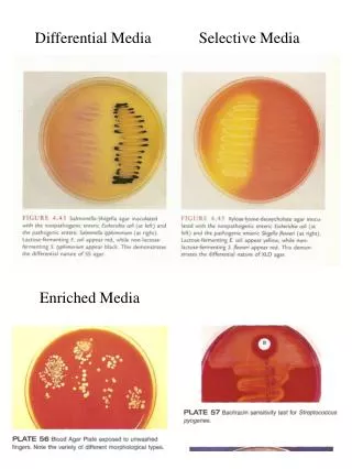
Differential media • Differential media does not necessarily inhibit bacterial growth, but instead makes the bacteria look different • Differential media works best with closely related organisms, and the differential agent is what causes the bacteria look different • Owing to the presence of certain dyes or chemicals in the media, the organisms will produce characteristic changes or growth patterns that are used for identification or differentiation. • A variety of selective and differential media are used in medical, diagnostic and water pollution laboratories, and in food and dairy laboratories. Mohammed laqqan
Some media are both selective and differential, that is, they are able to select against the growth of certain organisms while the organisms that do grow may exhibit some differential growth characteristics. • Three of the more common selective and differential media are described below and will be used in the laboratory exercise. Mohammed laqqan
Mannitol Salt Agar (MSA) Mannitol salt agar is a selective medium used for the isolation of pathogenic staphylococci. • The medium contains mannitol, a phenol red indicator, and 7.5% sodium chloride. • Ferment mannitol (differential): • Phenol red = pH indicator • Pink : pH >7.4 (basic) • Orange: pH = 7 (neutral) • Yellow: pH <6.8 (acidic) • Selective: The high salt concentration inhibits the growth of most bacteria other than staphylococci. • Note:The high salt content does not kill Gram negative bacteria, it just inhibits growth. Mohammed laqqan
On MSA, pathogenic Staphylococcus aureusproduces small colonies surrounded by yellow zones. The reason for this change in color is that S. aureus ferments the mannitol, producing an acid, which, in turn, changes the indicator from red to yellow. • Other Staphylococcus don’t ferment mannitol don’t produce a color change from the normal red-pink color of the medium. • The growth of other types of bacteria is generally inhibited. • S. aureus = salt tolerant; ferments mannitol (yellow) • S. epidermidis = salt tolerant; does NOT ferment (red) • M. luteus = not salt tolerant; does NOT grow on mannitol S. epidermidis M. luteus S. aureus Mohammed laqqan
EOSIN METHYLENE BLUE AGAR (EMB agar) • EMB is an undefined selective/differential medium. It contains aniline dyes (methylene blue and eosin), which inhibit the growth of Gram-positive bacteria selecting for Gram-negative bacteria. • EMB also contains lactose which makes the media differential based on an organisms ability to ferment lactose. • Sucrose is also included in the medium because certain members of the Enterobacteria or coliform group ferment sucrose more readily than they ferment lactose. These sugars provide favorable conditions for the growth of fecal coliforms. Mohammed laqqan
Lactose and sucrose fermenters will grow as dark colonies accompanied by a metallic green sheen (E. coli). • Organisms that slowly ferment lactose will appear as pink colonies (Enterobacter aerogenes are usually mucoid and much larger than colonies of E. coli). • Non-fermenters lactose or sucrose will remain colorless or take on the color of the medium such as Salmonella (one of the causative agents of food poisoning). • Note: This media is used to confirm the presence of E. coli in water samples contaminated with sewage or fecal material. • You will use this agar again with in the water sampling experiment to differentiate between E. coli and Enterobacter spp] Mohammed laqqan
The dark colonies produced on EMB agar is a result of the acid produced during lactose or sucrose fermentation precipitating the dyes in the media. K. pneumoniae produces dark colonies on EMB agar. Mohammed laqqan
EMB agar Mohammed laqqan
MacConkey AGAR • Used to isolate Gram negative enteric (coliforms) • Selective and differential medium. • Selective - Gram positive bacteria are inhibited by the presence of bile salts and crystal violet inhibitors in the medium. Most of gram negative bacteria will grow. • Differentiate- Between Gram negative bacteria by their ability to ferment lactose. • Pink colonies- Bacteria that ferment lactose ( These reactions are due to the acid produced by the fermentation of lactose. The acid end-products act on bile salts, and neutral red is absorbed by the precipitated salts. ). • Pale colonies- Non fermenters are no colored and transparent. • (Note: The Gram positive bacteria do not die on this media, their growth is just inhibited) Mohammed laqqan
MacConkey AGAR Mohammed laqqan
MAC agar Mohammed laqqan
Blood agar • Nutrient agar with 5% sheep blood • Cultivation of fastidious and non fastidious bacteria. • This media is differential because: • Certain bacteria produce enzymes hemolysins (exotoxin) that act on the red cells to produce either: • Beta hemolysis: Enzymes lyse the blood cells completely, producing a clear area around the colony. • Alpha hemolysis: Incomplete hemolysis produces a greenish discoloration around the colony. • Gamma hemolysis: No effect on the red cells. Mohammed laqqan
Procedure • Cultures of: • Staphylococcus aureus • Staphylococcus epidermidis • Escherichia coli • Enterobacter aerogenes • Salmonella enteritidis • Petri dishes of Mannitol Salt agar, EMB agar, MacConkey’s agar and nutrient agar • Procedure: • Divide each plate of MSA and MacConkey’s agar into two sections. Divide the • EMB agar plate into three sections. • Inoculate the MSA plate with S. aureus and S. epidermidis. • Inoculate MacConkey’s agar with E. coli and Salmonella enteritidis. • Inoculate the 3-sectional EMB agar plate with E. coli, Enterobacter aerogenes, and Salmonella enteritidis. • Divide one plate of nutrient agar into two sections and one into three sections. Inoculate the 2-sectional plate with the two species of Staphylococcus; inoculate the 3-sectional plate with the remaining three organisms. • Incubate all plates at 37°C for 48 hours. Mohammed laqqan
Observe the growth and appearance of colonies on all plates. • Notice that nutrient agar is neither a selective nor differential medium. Mohammed laqqan
End of lecture Mohammed laqqan