Download
1 / 23
320 likes | 770 Views
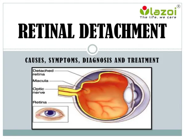
Retinal Detachment. Failure to diagnose retinal detachment is the second most common cause of large liability claims involving optometrists.

E N D
Failure to diagnose retinal detachment is the second most common cause of large liability claims involving optometrists.
If a patient presents with symptoms indicative of retinal detachment, or if a reasonable optometrist would suspect that the symptoms indicate the risk of a retinal detachment, a dilated fundus examination must be performed.
An elderly woman who had been the long-term patient of an optometrist complained of suddenly seeing a “spot” in the field of vision of one eye. The optometrist found acuity in the eye to be 20/20 and observed no pathology when examining the posterior pole through an undilated pupil. He told the patient she had a PVD and described the symptoms of retinal detachment. About 4 months later she suddenly began seeing light flashes in the eye, and a retinal exam revealed detachment affecting the macula.
For asymptomatic patients who are at risk for retinal detachment, a dilated fundus examination should be performed at the first examination and periodically thereafter. Common risk factors include: • history of past retinal detachment • myopia (-6 diopters or more) • open-angle glaucoma in myopic patients treated with strong miotic agents • aphakia or pseudophakia (especially the 6 months following cataract surgery), and capsulotomy (especially the 6 months following the laser procedure) • lattice degeneration • blunt ocular trauma (especially the year following the trauma) • proliferative retinopathies (e.g., proliferative stage of diabetes, sickle cell, branch retinal vein occlusion) • retinal detachment in fellow eye
History ofprevious detachment—if the detachment is from a non-traumatic cause, there may be a significant riskof detachment inthe fellow eye. Demarcation lines (below) indicate there is slowly progressive detachment
High myopia—the thin, stretched retina of high myopic eyes is at risk for tears (greater than 5 diopters 2% risk, greater than 10 diopters, 5% risk).
A severely myopic man (-18 D) complained of seeing numerous small spots and specks in one eye. Best acuity was 20/400, which the optometrist attributed to refractive amblyopia. Fundus exam was through an undilated pupil and was unremarkable. No information was provided concerning the symptoms of retinal detachment or the need for a follow-up examination. About 2 weeks after the exam acuity in the eye was reduced to hand motion. Evaluation revealed a retinal detachment, which had been present for more than the 2 weeks. A retinal tear was also found in the fellow eye (-7 D).
Pseudophakia—cataract surgery results in detachment in up to 2% of cases, depending on the technique used.The 6 monthsfollowing surgeryis when the eyeis most at risk.YAG capsulotomyalso creates up to a 3% risk of detachment.
Lattice degeneration—is thinning of the retina with liquified vitreous above the retina and adhesion to the retina at the lattice margins, which can result in detachment if there are holes at the latticemargins or a tear caused by posterior vitreousdetachment.
Open-angle glaucoma treated with strong miotics—high drug concentrations (6% pilocarpine) may cause intense miosis that leads to retinal stretching in high myopic eyes and a resultant tear.
Blunt trauma—the most common detachment is retinal dialysis, which occurs at the ora serrata and is slowly progressive, taking an average of 4 months to involve the macula.
Proliferative retinopathy—diabetes, vein occlusion, sickle cell, and other conditions cause neovascularization, which results in bleeding, fibrosis, and ultimately a tractional retinal detachment.
For purposes of litigation, the most important type of detachment is acute onset, symptomatic posterior vitreous detachment.
Patients with an acute onset, symptomatic posterior vitreous detachment (PVD) require a dilated fundus examination, because between 8% and 15% of cases have a retinal tear; of these, approximately one-third will develop into a retinal detachment.
Retinal detachment may occur (from an incompletely separated PVD) even though no tear is found at the time of examination; the 2 months following the onset of symptoms is the most likely period for a detachment to occur.
If a patient with acute onset, symptomatic PVD does not have a tear or detachment at the time of examination, a warning describing the symptoms of detachment must be given and the patient scheduled for a follow-up examination in 2 to 4weeks. Patients should not be dismissed from care until full separation of the vitreous has occurred(which can be as longas several months).
A finding of vitreous hemorrhage with symptoms of acute PVD indicates there is a 70% to 80% likelihood of an underlying retinal break.Referral for retinalevaluation is necessary.
All warnings should be noted in the patient record or documented through the use of a signed form.Handwritten entries do not need to be lengthy, but must describe the risk (“warned patient of the signs and symptoms of retinal detachment”), what to do if the risk occurs (“patient to RTC immediately for DFE if S & S occur”), and the consent obtained (“patient understood and agreed”).
A military retiree in his 60s was examined by the base OD because of the acute onset of “spots” in one eye. A dilated fundus exam with BIO revealed PVD. Seven weeks after the exam while climbing a ladder the man experienced a bright flash in the eye. He called and made an appointment, but for 6 days later. Retinal detachment involving the macula was found. A lawsuit was filed, alleging negligence and breach of informed consent, with the patient averring he had not been warned of the symptoms of detachment. At the trial, the doctor’s records were the key: he had written “PVD. Reassure. RTC PRN”.
Note how the doctor is trapped into admitting that telling the patient about the symptoms of retinal detachment was necessary if the PVD was incomplete, and that failure to bring the patient back in 2-4 weeks breaches the standard of care in such a situation.
CLAP Traps • The record must reflect whether the PVD is fully separated • If not separated, warnings and rescheduling are required • If fully separated, make it clear that any warning to the patient about the symptoms of detachment applies to a future PVD in the fellow eye
Run out of ideas for presents? Here’s something to give that special somebody for the holidays…