Download
1 / 41
590 likes | 2.13k Views
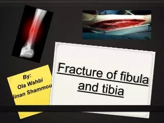
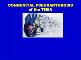
CONGENITAL PSEUDARTHROSIS of the TIBIA. ICLL: CPT. Pathology. Fibrous Hamartoma surrounding bone Associated with NF in over 50% No NF at CPT site. Where is the disease? Bone vs Periosteum. Observation: Slow bone remodelling of pin holes Remodelling is a periosteal function

E N D
CONGENITAL PSEUDARTHROSIS of the TIBIA ICLL: CPT
Pathology • Fibrous Hamartoma surrounding bone • Associated with NF in over 50% • No NF at CPT site
Where is the disease?Bone vs Periosteum Observation: Slow bone remodelling of pin holes Remodelling is a periosteal function New theory (Paley 1995: CPT is a periosteal disease and not a bone disease (the bone is affected secondarily)
Hermanns-Sachweh B et al: Vascular Changes in the Periosteum of Congenital Pseudarthrosis of the Tibia. Pathology Research and Practice 201, 305-312, 2005. • Neural cells forming a tight sheath around the periosteal arteries reminiscent of the way Schwann cells surround peripheral nerve fibers. • Result: accumulation of nerve cells in the periosteum cause narrowing or obliteration of the periosteal vessels. • Result: periosteum undergoes hypoxemic degeneration resulting in the formation of a thick fibrous cuff. This leads to impaired oxygen and nutrient supply to the subperiosteal bone with secondary atrophic bone changes.
Is this theory really new? • Codivilla (Italy) 1903 autogenous osteoperiosteal graft • Cambras (Cuba) 1977 periosteal graft from mother
CPT: Classifications • • Camurati 1930 • • Badgley 1952 • • Boyd 1958 • • Apoil 1970 • • Andersen 1973 • • Crawford 1986 • • Crawford 1999 • El-Rosassy-Paley 2000
I II III El-Rosassy-Paley Classification
Type I • Atrophic, narrow bone ends • Mobile • No previous surgery El-Rosassy-Paley Classification
Type II • Atrophic, narrow bone ends • Mobile • Previous surgery El-Rosassy-Paley Classification
Type III • Hypertrophic, wide bone ends • Stiff • Previous surgery El-Rosassy-Paley Classification
Type I Treatment Lateral AP
preop postop
3 year follow-up Flexible IM nail
Fassier-Duval Telescopic Nail
Type II Treatment Bone transport Acute shortening
acute shortening re-lengthening
bone transport healed
ReFx IM rod follow-up
Treatment • Type III • Closed distraction + compression • Gradual deformity correction • ± Lengthening • ± IM Rod (late)
distraction of CPT follow-up
pre-op compression healed
Treatment • Type III • Closed distraction • Gradual deformity correction • ± Lengthening • ± IM Rod (late)
distraction of CPT follow-up
RESULTS Periosteal & Bone Grafting with IM rod and Ilizarov 1997-2008 20 patients treated Union: 20/20
Don’t Forget Bracing until Skeletal Maturity