Download
1 / 21
210 likes | 240 Views
Explore the intricate anatomy and crucial functions of the cerebellum, the silent brain that plays a vital role in motor activity control and coordination. Learn about its physiological and anatomical divisions, input and output pathways, and the role it plays in coordinating movements.

E N D
1. Have u seen cranestanding on single leg ?2.A man while crossing the road sees a car coming but can not predict the speed of the car and is hit.3. A baller intendeding to throw a Yolker of his first ball to the batsman, hits the head of the keeper ?
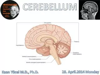
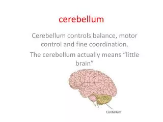
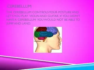
Cerebellum • Largest Part of the hind brain . • Surface area is 75% of the Cerebrum • BUT 10% of the weight of Cerebrum • Silent Brain • ( Can neither Perceive nor initiate sensory or motor activity) • Control motor functions in association with Cerebral cortex and basal ganglia , Regulator of the Motor Activity
Anatomy: Vermis: Narrow worm like central region(Nine Subdivisions) Cerebellar Hemispheres: • Two on both sides of vermis Divided in to: • Three lobes separated by 2 horizontal fissures Primary Fissure
Physiological Divisions Longitudinal imaginary line divides Cerebellar Hemispheres Lateral & intermediate zone Contd….
Physiological Subdivision of Cerebellum 1. Vestibulo-Cerebellum:Flocculonodolar Lobe Control of Equilibrium,Posture & Eye movements 2. Spino-Serebellum:Vermis + intermediate zones Topographical representation of muscle groups .Vermis - Axial & Proximal limb movements.(Connected with medial motor .Intermediate Zone -Distal Limb Movements ( connected with Lat.motor system) Concerned with Coordination of Movements 3. Cerebrocerebellum:Lateral Zone Connected with cerebral cortex Bilaterally. Mainly concerned with planning & Programming of sequential movements
Homunculus in CerebellumTopographical Representation of the Body • Vermis: • Movements of • Trunk • Shoulders • Neck • Hips • Intermediate Zone: • Movements of Limbs: • Especially Hands, Fingers, • Feet ,Toes • & Face • NO Body Parts on Lateral Zone Contd….
Cerebellum Input Signals: • From respective body parts • Corresponding topographical areas of motor cortex Output Signals: • Same areas of motor cortex • Red nucleus • Reticular formation Contd….
Cerebellum A. Input Pathways from Brain passes through: Three peduncles: Superior cerebellar peduncle : Midbrain Middle cerebellar peduncle : Pons Inferior cerebellar peduncle : Medulla i) Cortico-Ponto-Cerebellar pathway: Origin: Primary motor cortex Premotor cortex (Sensory cortex) To: Pontine nuclei Through: Middle cerebellar peduncle To: Contralateral cerebellar hemisphere Contd….
Cerebellum ii) Olivocerebellar Tract: Proprioceptors from the whole body Via Inferior olive Inferior cerebellar peduncle. all partsof cerebellum Inferior OliveReceives information from: Motor Cortex Basal Ganglia Reticular Formation Spinal Cord Contd….
iii) Vestibulocerebellar Tract: Vestibular nuclei (+ vestibular apparatus) Inferior peduncle Flocculonodular lobe + Fastigial nuclei iv) Reticulocerebellar Tract: Reticular formation Inferior peduncle Vermis V)Cuneocerebellar - from Head & Neck VI) Tectocerebellar - Visual & Auditatry
B. Input Pathways From Periphery (Proprioceptors) Two Ventral + two Dorsal spino cerebellar tracts i)Doral spino cerebellar tracts: Sensory information from Proprioceptors: Muscle Spindle, Golgi tendon organs, joint receptors, Tactile receptors of skin spinal cord (Klark’s Column) Ascend on the same side inferior cereb. Peduncle vermis + intermediate zone Klark’s Column extends from T1 to L3 2. Ventral Spino cerebellar tracts: Corticospinal tract + Rubrospinal tract & other motor fibers Ant. Horn of cord- Internal pattern Generators in cord (Take Efference Copy) >Cross to opposite side & Ascend up to mid brain. Cross 2nd time & enter via Sup. Cerebellar Peduncle to Vermis & Intermediate zone as above
Afferents to Cerebellum Image
Efferent Pathways From Cerebellum • Outer: Grey matter (cortex) • Inner: White matter Deep cerebellar nuclei: Groups of nerve cell bodies embedded in white matter • Fastigial nucleus • Interposed nucleus (Globose & Emboliform Nuclei • Dentate nucleus All out put signal originate (pass through) deep nuclei brain
Efferents from Cerebellum Image Note: No efferents through Middle cerebellar peduncle
Three Major Output Pathways 1. Vermis Fastigial nuclei Medulla & Pons Function: • + Vestibular Nuclei Equlibrium • + Reticular formation Postural attitudes • Intermediate Zone Interposed nucleus • Thalamus Cerebral cortex • Thalamus Basal Ganglia • Red nucleus & reticular formation Function: Coordination and reciprocal contractions of agonist and antagonist muscles (esp. hands and fingers) Contd….
Lateral Zone of Cerebellum Dentate nucleus Thalamus Cerebral Cortex Function: No direct input from peripheral body parts • Two-way communication with cerebral cortex and basal ganglia • Planning & coordination of sequential movements , Smooth Progression Orderly sequence • Timing Function Time the sequential movements when to start and stop • Extramotor predictive function Can predict from changing visual scenes how rapidly a person is approaching an object
Functional Circuits of Cerebellum • About 30 million functional units: • Three Layers of the Cerebellar Cortex: 1. Molecular Layer 2. Purkinjee Cell Layer 3. Granule Cell Layer TWO major Afferent (Incoming) Fibers: a. Climbing Fibers b. Mossy Fibers -
Functional Neuronal Circuit of Cerebellum P - + Inf. Olive