Download
1 / 44
450 likes | 670 Views
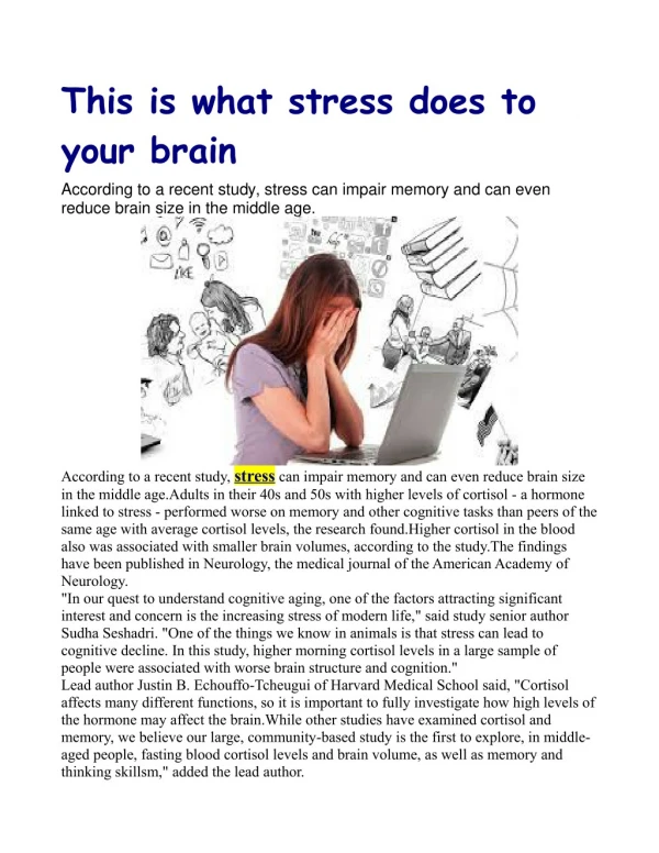
STRESS, GLUCOCORTICOIDS, AND YOUR BRAIN. Stress Hormones: Glucocorticoids. Cortisol in the human Corticosterone in the rat Secreted by the cortex of the adrenal gland. Landfield et al .(1979,1981). Basal (resting) levels of glucocorticoids ( GCs ) rise with age in rats

E N D
Stress Hormones: Glucocorticoids • Cortisol in the human • Corticosterone in the rat • Secreted by the cortex of the adrenal gland
Landfield etal.(1979,1981) • Basal (resting) levels of glucocorticoids (GCs) rise with age in rats • The greater the basal level of GCs in old rats, the greater the cell loss in the hippocampus
Landfield etal.- cont. Middle Aged rats (12 months) Removed the adrenal glands (Adx) Replacement injections of GCs (low concentration for life -1.0 yr) Two years old No cell loss in hippocampus
Landfieldetal. - results No. of hippo. neurons per 100 sq. microns of tissue
Sapolsky etal. (1985 ff.) • Young rats (3-5 months) • Daily injections of stress levels of GCs - Two weeks (acute) or - Three months (chronic) RESULTS - acute treatment No hippocampal cell loss - chronic treatment significant hippocampal cell loss*
What natural conditions contribute to increased glucocorticoid levels?
Sapolsky etal. (1989) • Field studies - male baboons in Kenya • Measured basal levels of GCs (cortisol) - 6 years • determined social dominance rank • RESULTS: - lowest ranking males subjected to unpredictable stress - lowest ranking males have highest GC levels*
Uno etal.(1989). Hippocampal Damage Associated with Prolonged and Fatal Stress in Primates.
Uno etal. (1989) • Observations under natural conditions • Eight vervet monkeys • Agricultural pests primate center in Kenya • All died suddenly over two year period • Control group (n=5) of euthanized animals from the center • Autopsies upon death • RESULTS: - all eight from sudden death group had gastric ulcers
Uno et al. (1989) - cont. • Enlarged adrenal gland cortex in each SD monkey • Enhanced numbers of bite scars on SD monkeys • Shrunken cells in hippocampus of all SD monkeys • Reduced numbers of cells in various cell layers of the hippocanpus of males* - pyramidal neurons in areas CA1, CA3, CA4 Conclude: - SD animals subjected to prolonged stress - Prolonged stress increased GC secretion - Increased GC secretion hippocampal damage
Sapolsky etal. (1990) - Hippocampal Damage Associated with Prolonged Glucocorticoid Exposure in Primates
Sapolsky et al. (1990) • Four vervet monkeys • Cortisol pellets implanted into hippo. on one side • Cholesterol(control) pellet into hippo. on other side • One year later examine hippo. for pathology RESULTS: - soma shrinkage on cortisol side* - dendritic atrophy on cortisol side
Summary • GC basal levels increase with age in rats • GCs can cause hippocampal pathology in rats and primates
How do glucocorticoids damage hippocampal neurons? • They endanger neurons by increasing their vulnerability to metabolic insults • Metabolic insults - hypoglycemia - hypoxia-ischemia • GCs inhibit glucose transport into hippocampal neurons Cell Death Energy Depletion Additional Energy Depletion
What role does stress play in hippocampal cell damage during metabolic insults?
Stein-Behrens etal.(1994).Stress Exacerbates Cell Loss in the Hippocampus.
Stein-Behrens etal. (1994) • Metabolic insult Kainic acid • Kainic acid Glutamic acid agonist • Kainic acid excites neurons depletes energy EXPERIMENT 1: • Five groups of ADX rats 1. 100% GCs 2. 60% GCs 3. 15% GCs Replacement injections for 4. 0% GCs 3 days 5. Stress GC level
Stein-Behrens etal. – cont. • Inject Kainic acid into the hippocampus • Euthanize 3 hours later • Examine neuron damage in the hippocampus • RESULTS: - Increasing amounts of neuron damage with increasing doses of GCs*
Will stress exacerbate hippocampal damage produced by Kainic acid?
Stein-Behrens etal. – cont. EXPERIMENT 2: • Normal rats, no ADX • Subject to three days of different stressors (cold, restraint, mix social grps.) • Inject Kainic acid into the hippocampus • Euthanize three hours later • Examine neuron damage in the hippocampus RESULTS: - Stress exacerbated kainic acid damage* High levels of GCs
What is the behavioral significance of increased levels of GCs? • REMEMBER: Landfield’s results - AGED RATS HIGH GC LEVELS - AGED RATS HIPPO. CELL LOSS • Hippocampal damage Memory deficits
Do aged rats with high glucocorticoid levels show hippocampal pathology and memory impairments?
Issa etal. (1990). Hypothalamic-pituitary-adrenal activity in aged, cognitively impaired and cognitively unimpaired rats
Issa etal. (1990) • Aged rats: 23-27 mos. • Young controls: 6-7 mos. • Behavioral screening in Morris Water maze 1. N=58 old rats 2. N=19 young rats • Hippocampal cell counts • Basal and stress-induced GC levels • GC binding in hippocampus
Issa etal.(1990) - cont. • RESULTS: Behavioral screening*: Started with 58 old rats 1. 16 (28%) = aged impaired 2. 20 (34%) = aged unimpaired 3. 22 (38%) = in between and excluded Young control rats Hippocampal cell counts: Control > aged unimpaired > aged impaired*
Issa etal.- cont. • Basal and stress GC levels*: - basal: Aged impaired secrete higher levels than aged unimpaired or young rats - stress: Aged impaired secrete higher levels than aged unimpaired or young rats • GC binding in hippocampus*: Aged impaired rats show less binding than other two groups
What are the implications of this research for humans? Non-elderly human populations with high GC levels -Depressed patients - Cushing’s syndrome Elderly humans – some have high cortisol levels
Sheline etal. (1996, 1999). Hippocampal atrophy in recurrent major depression.
Sheline etal. • Magnetic resonance imaging (MRI) • Humans with a prolonged history of depression • Image taken while patients were no longer depressed • RESULTS: - Left and right hippocampus reduced in volume compared to control group* - The longer the time depressed, the less the hippocampal volume*
Starkman etal. (1992). Hippocampal formation volume, memory dysfunction, and cortisol levels in patients with Cushing’s syndrome.
Starkman et al.(1992) • Magnetic resonance imaging • Memory tests • Cortisol levels RESULTS: • The greater the cortisol level, the less the hippocampal volume* • Cushing’s syndrome patients demonstrate memory impairments - the less the hippocampal volume, the greater the memory impairment
Lupien etal. (1998). Cortisol levels during human aging predict hippocampal atrophy and memory deficits.
Lupien etal. (1998) • Measured cortisol levels in the elderly for 5-6 years • Two groups: 1. increasing/high levels of cortisol 2. decreasing/moderate levels of cortisol • Memory test: - line drawings - immediate and delayed (24 hr.) recall
Lupien etal.-cont. • RESULTS: - increasing/high group delayed memory deficit - increasing/high group decreased hippo.volume
Post Traumatic Stress Disorder (PTSD) • Combat veterans, victims of rape, childhood sexual abuse, accidents, violent crimes • Symptoms: - flashbacks - nightmares - sleep disorders - emotional disorders - enhanced startle reflex - disorders of memory and concentration
Bremner etal. (1995). MRI-based measurement of hippocampal volume in patients with combat-related posttraumatic stress disorder
Bremner etal.(1995) • Subjects: - 26 Vietnam combat veterans with diagnosed PTSD - 22 matched controls (age, handedness, sex, race, age, weight, height, SES, yrs. education, alcohol abuse) • MRI • Assessed verbal memory (Wechsler Memory Scale)
Bremner etal.(1995) • RESULTS: - Right hippocampal volume is reduced by 8% in PTSDs* - Reduction selective to the hippocampus - PTSDs demonstrated memory impairments - The less the volume of the right hippocampus, the worse the memory
Bremner etal.(1997). Magnetic resonance imaging-based measurement of hippocampal volume in postraumatic stress disorder related to childhood physical and sexual abuse – a preliminary study
Bremner etal.(1997) • 17 adult survivors of severe childhood physical/sexual abuse diagnosed with PTSD • 17 matched controls • MRI • Assessed verbal memory RESULTS: - Left hippocampal volume is reduced by 12% in PTSDs - Reduction is selective to the hippocampus - Impaired verbal memory - The longer the history of abuse, the less the hippocampal volume
Important Question • What brain circuits are activated by sustained stress to cause hypersecretion of cortisol?
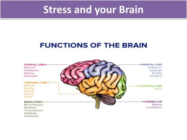
Fear of the bear Activates the amygdala Activates the hypothalamus (Paraventricular nucleus) Releases CRF into the anterior lobe of the pituitary gland Releases ACTH Adrenal gland glucocorticoids