Download

1 / 45
460 likes | 746 Views
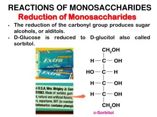
The two families of monosaccharides are aldose and ketoses. Monosaccharides are colorless, crystalline solids that are freely soluble in water but insoluble in nonpolar solvents. Most have a sweet taste. The backbone of common monosaccharide are unbranched carbon chain.

E N D
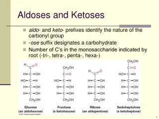
The two families of monosaccharides are aldose and ketoses • Monosaccharides are colorless, crystalline solids that are freely soluble in water but insoluble in nonpolar solvents. Most have a sweet taste. The backbone of common monosaccharide are unbranched carbon chain. • Aldose Vs. Ketose: In the open-chains form, (carbonyl group) is at end of the carbon chain. Trioses, tetroses, pentoses, hexoses, and heptoses (aldohexose D-glucose and ketoheose D-fructose
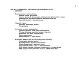
Monosaccharies have asymmetric centers • A molecule with n chiral center can have 2n stereoisomers. • Chiral center most distant from the carbonyl carbon—define D, L isomer (refer to D-glyceraldehyde)--- most of the hexoses of living organisms are D isomers. • Ketose—insertion of ul into the name of a corresponding aldose; D-ribulose = aldopentose D-ribose (ketopentose).
Epimers • Epimers: Two sugars that different only in the configuration around one carbon atom. • D-glucose and D-mannose, which differ only in the stereochemistry at C-2, are epimers, as are D-glucose and D-galactose (which differ at C-4)
Formation of hemiacetals and hemiketals • An aldehyde or keton can react with an alcohol in a 1:1 ration to yield a hemiacetal or hemiketal, creating a new chiral center at the carbonyl carbon. • Substitution of a second alcohol molecule produce an acetal or ketal. When the second alcohol is part of another sugar molecule, the can produced is a glycosidic bond
Common monosaccharides have cyclic structures: Formation of the two cyclic forms of D-glucose • Reaction between the aldehyde group at C-1 and the hydroxyl group at C-5 forms a hemiacetal linkage, producing either of two steroisomers, the a andbanomers (differ in anomeric carbon = hemiacetal or hemiketal), which differ only in the sterochemistry around the hemiacetal carbon. • The introversion of a (1/3) and b (2/3) anomer is called mutarotation (identical optic properties). glycosidic bond
Pyranoses and Furanoses • The pyranose forms of D-glucose and the furanose forms of D-fructose • Aldohexoses also exist in cyclic forms having five-membered rings, The six-membered aldopyranose ring is much more stable than the aldofuranose ring and predominated in aldohexose solution. Only aldoses having five or more carbon atoms can form pyroanose rings.
Conformation formulas of pyranoses chair boat • Constituents on the ring carbons may be either axial (ax), projection parallel with the vertical axis through the ring or equatorial (eq), projecting roughly perpendicular to this axis. Generally, constituents in the equatorial positions are less sterically hindered by neighboring constituents, and the conformations with their bulky constituents in equatorial positions are favored. • Boat– is only seen in derivatives with very bulk constituents
The acidic sugars contain a carboxylate--- confer a negative charge at neutral pH. D-glucono-d-lactone – formation of ester linkage between the C-1 and C-5. Amino sugar—NH2 is replaced –OH. deoxy sugar--substitution of –H for –OH. The deoxy sugars of nature as the L isomers Some hexose derivatives important in biology
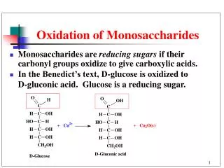
Monosaccharides are reducing agents - Fehling’s reaction • Reducing sugars: glucose and other sugars capable of reducing ferric or cupric ion (carbonyl carbon is oxidized to a carboxyl group (Fe3+ , and Cu2+ to Fe2+ and Cu+ --- red cuprous oxide precipitate).
Dissaccharides contain a glycosidic bond • Disaccharides consist of two mono-saccharide joined covalently by an O-glycosidic bond formed when a glydroxyl of one sugar reacts with the anomeric carbon of the other. • Sugar (sucrose) containing the anomeric carbon atom cannot exist in linear form and no longer acts as a reducing sugar. • Nonreducing disaccharides are named as glycosides
Disaccharides – a Vs b glycosidic linkage • Disaccharides (such as maltose, lactose, and sucrose) consist of two monosaccharides joined covalently by an O-glycosidic bond, which is formed when a hydroxyl group of one sugar reacts with the anomeric carbon of the other. • Glycosidic bonds are readily hydrolyzed by acid but resist cleavage by base. • the name describes the compound with its nonreducing end to the left, and we can “build up” the name in the following order. (1) Give the configuration (or ) at the anomeric carbon joining the first monosaccharide unit (on the left) to the second. (2) Name the nonreducing residue; to distinguish five- and six-membered ring structures, insert “furano” or “pyrano” into the name. (3) Indicate in parentheses the two carbon atoms joined by the glycosidic bond, with an arrow connecting the two numbers; (4) Name the second residue. If there is a third residue, describe the second glycosidic bond by the same conventions. ?
Polysaccharides--glycans • may compose of one, two, or several different monosaccharide, in straight or branched chains of varying length • Homo- vs. hetero-polysacchairdes. • As fuel or structure element
Starch and glycogen granules • Polysaccharides do not have definite molecular weight. (protein is on the template of defined sequence and length; no template of polysaccharides) • Starch– amylose (long and unbranched chains of glucose) and amylopectin (branched 24 to 30). • Glycogen — more extensively branched and more compact than starch. • Dextransare bacterial and yeast polysaccharides made up of (1 - 6)-linked poly-D-glucose; all have (1 - 3) branches, and some also have (1 - 2) or (1 - 4) branches. Dental plaque, formed by bacteria growing on the surface of teeth, is rich in dextrans.
The structure of cellulose • b1-4 linkage –most stable conformation for the polymer is that in which each chair is turned 180o relative to its neighbors, yielding a straight, extended chain. (inter and intra H bonds)--- water can not get in. • Digested by cellulase (termites, fungi, bacteria, ruminants)
Cellulose breakdown by wood fungi • All wood fungi have the enzyme cellulase, which breaks the (1- 4) glycosidic bonds in cellulose, such that wood is a source of metabolizable sugar (glucose) for the fungus. • The only vertebrates able to use cellulose as food are cattle and other ruminants (sheep, goats, camels, giraffes). The extra stomach compartment (rumen) of a ruminant teems with bacteria (symbiotic microorganism, Trichonympha) and protists that secrete cellulase
Chitin — polymer of N-acetylglucosamine in b linkage • is a linear homopolysaccharide composed of N-acetylglucosamine residues in linkage • Indigested by most vertebrate animal. • Exoskeletons of arthropods—insects, lobsters, and crabs.
Conformation at the glycosidic bonds of cellulose, amylose and dextran • The three-dimensional structures of these molecules can be described in terms of the dihedral (由兩個平面構成的) angles, and , made with the glycosidic bond. • Cellulose, the most stable conformation is that in which each chair is turned 180 relative to its neighbors, yielding a straight, extended chain. • The most stable three-dimensional structure for starch and glycogen is a tightly coiled helix-Each residue along the amylose chain forms a 60 angle with the preceding residue, so the helical structure has six residues per turn.
A map of favored conformations for oligosaccharides and polysaccharides • The torsion angles which define the spatial relationship between adjacent rings, can in principle have any value from 0 to 360. In fact, some of the torsion angles would give conformations that are sterically hindered, whereas others give conformations that maximize hydrogen bonding. • analogous to the Ramachandran plot for peptides
The structure of starch (amylose) • In the most stable conformation of adjacent rigid chairs, the polysaccharide chain is curved, rather than linear. • The a1-4 linkage causes these polymers to assume tightly coiled helical structures (more compact). • Hydrolysis by a amylases (saliva and intestinal secretion)
Bacterial cell walls contain peptidoglycans proteoglycans • Polymer of N-actylglucosamide, cross-linked with short peptides • Lysozyme (tear, bacterial viruses)—lyses the (b1-4) glycosidic bonds. • Penicillin prevents synthesis of cross-links leaving the cell wall too weak to resist osmotic lysis.
The structure of agarose • The repeating unit consists of D-galactose (1- 4)-linked to 3,6-anhydro-L-galactose (in which an ether ring connects C-3 and C-6). These units are joined by (1- 3) glycosidic links to form a polymer 600 to 700 residues long. A small fraction of the 3,6-anhydrogalactose residues have a sulfate ester at C-2. • When a suspension of agarose in water is heated and cooled, the agarose forms a double helix: two molecules in parallel orientation twist together with a helix repeat of three residues; water molecules are trapped in the central cavity. These structures in turn associate with each other to form a gel—a three-dimensional matrix that traps large amounts of water.
Repeating units of some common glycosaminoglycans of extracellular matrix • extracellular matrix - a gel-like material that fill between multicellular organisms - composed of an interlocking meshwork of heteropolysaccharides and fibrous proteins: collagen, elastin, fibronectin, and laminin. • One is N-acetylglucosamine or galactosamine; the other is D-glucuronic (most cases) • Esterified with sulfate (negative charge)--- assume extended conformation in solution. Attaches to proteins– proteoglycans. pliability (易曲折;柔軟)
Glycosaminoglycans • Hyaluronic acid (Glass): lubricants in synovial fluid (關節滑液), eye, cartilage and tendons; hyaluronidase secreted by bacteria — bacteria invasion. Similar enzyme for sperm to penetrate ovum. • Chondroitin sulfate (Cartilage): tensile strength pf cartilage, tendons and ligament, aorta. • Dermatan sulfate (Skin): skin, blood vessel and heart valves. Pliability of skin. • Keratan sulfates (horn): cornea, horn, hair, hoof, nails, claws, no uronic acid. • Heparin (liver): made in mast cell- a anticoagulant with highest negative charge density, release to blood, inhibit blood clotting by binding to antithrombin III - bind to and inhibit thrombin, a protease essential to blood clotting.
Interaction between a glycosaminoglycan and its binding protein • Fibroblast growth factor (FGF1), its cell surface receptor (FGFR), and a short segment of a glycosaminoglycan (heparin) were co-crystallized: • red- predominantly negative charge; blue - predominantly positive charge. Heparin- negative charges (SO3- and COO-) attracted to the positive
Proteoglycan: macromolecules of cell surface or extracellular matrix in which one or more glycosaminoglycan chain are jointed covalently to a membrane protein or a secreted protein. Major components of cartilage. Glycoprotein: have one or several oligosaccharides of varying complexity joined covalently to a protein– outer surface plasma membrane, extracellular matrix and in the blood. Glycolipid: membrane lipid in which the hydrophilic head are oligosaccharides. A typical trisaccharide linker connects a glycosaminoglycan— ex. chondroitin sulfate (orange)— to a Ser residue (red) in the core protein. The xylose residue at the reducing end of the linker is joined by its anomeric carbon to the hydroxyl of the Ser residue. Proteoglycan structure, showing the trisaccharidebridge glycosaminoglycan
Proteoglycan structure of an integral membrane protein -- syndecan • A core protein of the plasma membrane. The N terminal on the extracellular side of the membrane is covalently attached to three heparan sulfate and two chondroitin sulfate chain. • S domains - highly sulfated domains alternate with domains having unmodified GlcNAc and GlcA residues (N-acetylated, or NA domains). - bind specifically to extracellular proteins and signaling molecules to alter their activities.
A proteoglycan aggregate of the extracellular matrix • One very long molecule of hyaluronate is associated noncovalently with about 100 molecules of the core protein aggrecan. Each aggrecan molecule contains many covalently bound chondroitin sulfate and keratan sulfate chains. Link proteins situated at the junction between each core protein and the hyaluronate backbone mediate the core protein–hyaluronate interaction.
Interactions between cells and extracellular matrix • The associating between cells and the proteoglycan of extracellular matrix is mediated by a membrane protein (integrin) and by an extracellular portein (fibronectin) with binding sites for both integrin and the proteoglycan
Oligosaccharide linkages in glycoproteins (secretion protein and cell surface) • O-linked oligosaccharides– glycosidic bond to hydroxyl group of Ser or Thr residues. • N-linked have and N-glycosyl bond to the amide nitrogen of an Asn • Alter polarity and solubility; protein folding, protect proteins from attack by proteolytic enzymes, increasing structural complexity • add in Golgi complex
Bacterial liposaccharides (glycolipid) • Ganglioside-membrane lipids of eukaryotic cells, the polar group is a complex oligosaccharide containing sialic acid (determine human blood) • Target of Ab. Serotype: strains that are distinguished on the basis of antigenic properties. • Toxic to human (lowered blood pressure toxic shock syndrome)--- Gram-negative bacteria infection.
Oligosaccharide - lectin interactions mediated many biological processes • Lectins:proteins that bind carbohydrates with high affinity and specificity (H bonds…) --- cell-cell interaction and adhesion. - useful reagents for detecting and separating glycoproteins with different oligosaccharide moieties. • Sialic acid residues situated at the ends of the oligosaccharide chains of many plasma glycoproteins — protect the proteins from uptake and degradation. • sialidase (neuraminidase) remove sialic acid – asialoglycoprotein receptors binds => triggers endocytosis and destruction of the protein, another i.e. RBC • The lectin of the influenza virus (HA) - binding of the virus to a sialic acid–containing oligosaccharide on the host surface, a viral sialidase removes the terminal sialic acid residue, triggering the entry of the virus into the cell. Inhibitors of this enzyme are used clinically in the treatment of influenza.
Role of lectin-ligand interactions in lymphocye movement to the site of and infection or injury • An infection site, P-selectin on the surface of capillary endothelial cells interacts with a specific oligosaccharide of the gluycoproteins of circulating T lymphocytes --- integrin interact with E-selectin (endothelial cell, L-selectin on the T cell) • Cholera toxin molecule entering intestinal cells (oligosaccharide of ganglioside GM1). • another i.e. Pertussis toxin
Helicobacter pylori adhering to the gastric surface • Helicobacter pylori (bacterial membrance lectin), adheres to the inner surface of the stomach (oligosaccharide) – O blood type Leb, synthesized analogs of the Leb. • Lectins also act intracellularly. An oligosaccharide containing mannose 6-phosphate marks newly synthesized proteins in the Golgi complex for transfer to the lysosome.
Hydrophobic interactions of sugar residues • Sugar units such as galactose have a more polar side (the top of the chair, with the ring oxygen and several hydroxyls), available to hydrogen-bond with the lectin, and a less polar side that can have hydrophobic interactions with nonpolar side chains in the protein, such as the indole ring of tryptophan.
Roles of oligosaccharides in recognition and adhesion at the cell surface (a) Oligosaccharides with unique structures (components of a variety of glycoproteins or glycolipids on the outer surface of plasma membranes, interact with high specificity and affinity with lectins in the extracellular milieu (周圍環境). (b) Viruses that infect animal cells, such as the influenza virus, bind to cell surface glycoproteins as the first step in infection. (c) Bacterial toxins, such as the cholera and pertussis toxins, bind to a surface glycolipid before entering a cell. (d) Some bacteria, such as H. pylori, adhere to and then colonize or infect animal cells. (e) Selectins (lectins) in the plasma membrane of certain cells mediate cell-cell interactions, such as those of T lymphocytes with the endothelial cells of the capillary wall at an infection site. (f) The mannose 6-phosphate receptor/lectin of the trans Golgi complex binds to the oligosaccharide of lysosomal enzymes, targeting them for transfer into the lysosome.