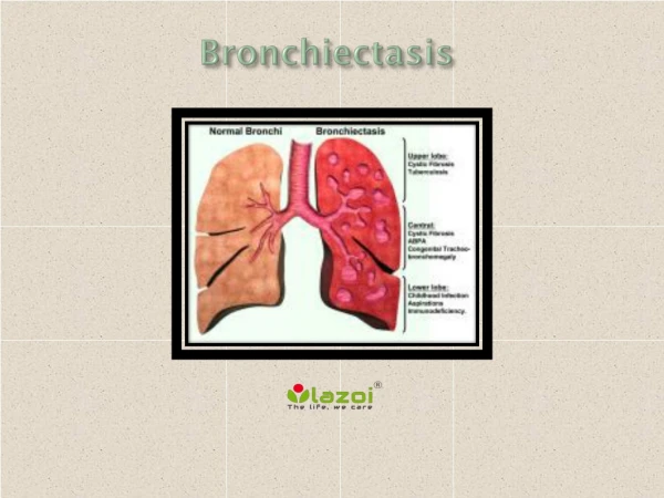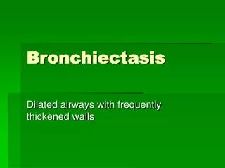
An interesting CAuSE OF bronchiectasis
An interesting CAuSE OF bronchiectasis. Dr.S.KADHIRVEL –VI MU CHIEF -Dr C .DHARMARAJ. 60Y /M presented with C/o cough with expectoration -2 weeks Copious yellow coloured sputum foul smelling ,not blood stained H/o breathlessness , Gr II

An interesting CAuSE OF bronchiectasis
E N D
Presentation Transcript
An interesting CAuSE OF bronchiectasis Dr.S.KADHIRVEL –VI MU CHIEF -Dr C .DHARMARAJ
60Y /M presented with • C/o cough with expectoration -2 weeks Copious yellow coloured sputum foul smelling ,not blood stained • H/o breathlessness ,Gr II • H/o chest pain ,pleuritic type over the right hemithorax • No h/o fever • H/o abdominal pain –dull dragging type over the right upper abdomen • No h/o palpitation
No h/o loose stools /bloody stools in recent past • No h/o decreased urine output • No h/o altered sensorium/seizures • No h/o blurring of vision
Past history: • K/c/o COPD on Rx for past few years. • K/c/o hydatid cyst – right lobe of liver,operated in a private hospital in 2015. • Not a k/c/o DM/SHT/PT/BA/CAD/seizures
Personal history: • Mixed diet • Chronic alcoholic 350ml -3 times a week for about 25 yrs • Chronic smoker -1-2 pack beedies for 35yrs • Normal bladder and bowel habits
General examination O/E Pt conscious Oriented , Mildly dyspnoeic febrile(+)-100.3F Pallor(+) clubbing –pandigital grade II No cyanosis No pedal edema No generalisedlymphadenopathy Hydration-good
Systemic examination • RS: Bilateral Air Entry(+), NVBS+ Trachea in midline Chest wall symmetrical, movements decreased in the right lower hemithorax B/L wheeze (+) –expiratory,poly phonic Crepts(mid inspiratory) –coarse leathery over the right mammary , infra axillary and lower inter scapular regions.
CVS:S1S2(+), No murmurs • P/A : soft ,Bowel Sounds(+) Liver enlarged(17cms) ,3cms below the right costal margin minimal tenderness in the right hypochondrium and epigastrium Spleen not palpable No free fluid. • CNS:No FND
Provisional diagnosis • COPD – infective exacerbation • Right middle lobe bronchiectasis • Right lower lobe pneumonia • ?Recurrent hydatid cyst –right lobe of liver
TREATMENT • NPO ,IVF • Injcefotaxime 1gm IV tds • Injmetronidazole 500mg iv tds • Injderiphylline 1amp iv tds • T.Ranitidine 150 mg bd • Salbutamolnebulisation 4th hourly • Abdominal girth chart • Vitals monitoring
USG-Abdomen • Multiple cystic lesions ,largest 12.1x10.7cms with daughter cysts and areas of wall calcifications noted in the right lobe of liver(seg 6&7) with wall calcifications • Other solid organs normal in size and echoes • No free fluid in abdomen and pelvis • No pleural effusion • s/o hydatid cyst recurrence –right lobe of liver.
Surgery opinion: decreased air entry –rt lower lobe Mild gaurding , tenderness –upper abdomen No rigidity P/R :normal Suggested: NPO IVF Xray chest and erect abdomen CT abdomen Abdominal girth chart SGE (o) and review
SGE opinion Suggested : • LFT • Repeat usg abdomen and pelvis • CT abdomen and pelvis USG review • No evidence of rupture of hydatid cyst at present
Thoracic physician opinion • Right middle lobe bronchiectasis • Suggested : sputum AFB HIV screening If isputum negative for AFB – plan FOB HIV screening – negative Sputum for AFB isnegative
Cardio thoracic surgeon opinion • CXR –no e/o hydro/pneumothorax • Suggested – CT chest and review
CT chest • E/o hydatid cyst in the lower part of the right middle lobe and upper part of the lower lobe • Measuring 10x12cms • Associated bronchiectatic changes present • E/o liver hydatid in segments 6 and 7 measuring 8x10 cmswith inflammatory changes noted. • No fluid in the abdomen /pelvis and pleural cavity
Cardio thoracic surgeon review • PFT • Sputum AFB and C&S • Repeat CXR • Planned for surgery -thoracotomy and enucleation • suggested : To add • Injampicillin 1gm iv bd • Injofloxacin 100mg iv bd • T.albendazole 400mg 1-0-1 • Chest physiotherapy • Vitals monitoring
PFT • Planned for surgery and assessed for anaesthetic fitness • Cardiac fitness was obtained • Echo showed Right atrium compressed by a large cystic lesion. IMPRESSION: SEVERE OBSTRUCTION
Pt developed sudden dyspnoea and hemodynamic instability • Shifted to CTS ICU and monitored • CT chest was taken and it showed right hydropneumothorax and partial collapse of the right lung and left side mediastinal shift s/o rupture of hydatid lung in to pleural space • Pt was stabilized and ICD tube inserted in the right 5thIntercostalspace • 400ml of dirty fluid -?pus drained
Operative findings • Diagnosis: ruptured hydatid middle lobe of right lung with liver hydatid • Procedure: excision of lung hydatid with removal of liver cyst. • Intra op findings:excision of lung hydatid -right middle lobe with removal of liver cyst through the diaphragm. Liver cavity obliterated . Diaphragm sutured and checked for air leaks. • Right lung adequately expanded after keeping suitable drains . Pt was shifted to IRCU and recovery monitored. 2 units of FFP and 1 packed cell unit were transfused.
To highlight the unusual cause of bronchiectasis • To discuss the manifestations and complications of hydatid disease • Synchronous involvement of liver and lung