Download
1 / 48
490 likes | 604 Views
Understand the physiological changes during pregnancy, the definition of postpartum hemorrhage, its severe effects, types, etiology, risk factors, prevention, and medical management to save lives.

E N D
Background • Physiologic changes during pregnancy • Increase in plasma volume by 40% • Increase red cell mass by 25% • Definition of postpartum hemorrhage • 500 mL after vaginal delivery • 1000 ml after cesarean delivery • Severe bleeding is #1 worldwide cause of maternal death • 140,000 women die each year from hemorrhage • 1 every 4 minutes • Other serious sequelae • coagulopathy, shock, loss of fertility • Hemorrhage frequently occurs without any warning
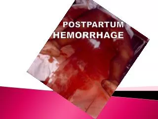
Definition: • Excessive blood loss during or following the 3rd stage of labor, more than 500 ml of blood loss following normal vaginal delivery of the fetus or 1000ml following Cesarean section. • Clinically the amount of blood loss from or into the genital tract which will adversely affect the general condition of the patient • Hemorrhage leading to fall in hematocrit by 10 %. • Incidence – 1- 4 % • Normally , the empty contracted uterus does not bleed.
Types 1] Primary 2] Secondary • Primary – bleeding occurs following delivery of the baby up to 24 hours • Primary is two types: A] Third Stage hemorrhage B] True Post Partum hemorrhage within 1st 24 hours. • Secondary : after 24hrs of delivery till the end of the pueperium .
Etiology – Primary Hemorrhage • Primary hemorrhage occurs in 1st 24 hours • Caused by The Four T’s • Tone – atony (80% of all cases) • Tissue – retained POC, accreta, uterine inversion • Trauma – cervical or vaginal laceration, rupture • Thrombic events – defects in coagulation • Inherited or acquired
PPH: UTERINE ATONY • Most dangerous • Uterus although empty, fail to contract and control bleeding from the placental site following the delivery of the placenta. • PREDISPOSING FACTORS • Over distention of uterus (multiple pregnancy, polyhydromnious, macrosomia) • Retained product of conception • Prolonged labour • Oxytocin augmentation of labour • Grandmultiparity • Antepartumhaemorrhage • Uterine fibroid • General anesthetic drugs (halothane) • Precipitate delivery • Chorioamnionitis • Magnesium sulphate treatment of PIH • Anemia
PPH: RETAINED PLACENTA • Defined as failure of the placenta to be expelled within 30 minutes after delivery of the fetus. • 2% of deliveries continues bleeding • Causes: – Placenta separated but undelivered – Placenta partly or wholly attached – Placenta accreta
PPH: GENITAL TRACT TRAUMA • Commonly follow an assisted delivery (forceps, ventouse) • Episiotomy can sometimes extends upwards and cause bleeding. • Uterine rupture at – previous caesarean section – previous myomectomy
PPH: Hematomas • occur when bleeding into a loose connective tissue occurs while overlying tissue remains intact. Blood collects 25-500 ml. in the soft tissue • Related to vascular injury during spontaneous or assisted delivery involving the vulva or vagina. • Vulvar hematoma – a discolored bulging mass producing deep ,severe, unrelieved pain with feelings of pressure. • Symptoms : • Severe vulvar pain • Unilateral purplish discoloration of the perineum or buttocks which are firm and tender. • Feeling fullness in the vagina Management: • Application of small ice packs • Surgical evacuation
Placental site 1- Atony 2- Retention of a partially or completed separated placenta or placental fragments 3- Hypofibrinogeamia or afibrinogeneamia • Extraplacental site - Laceration of the perineum , vulva , vaginal . Or cervix and rarely unrecognized rupture uterus
Risk Factors • Prolonged labor (also augmented labor) • Rapid labor • History of postpartum hemorrhage • Preeclampsia • Distended uterus • Macrosomia, twins, polyhydramnios • Chorioamnionitis • Operative delivery
Prevention • One key to prevention • One inexpensive intervention—active management of the third stage of labor (AMTSL)—could eliminate at least half of postpartum hemorrhage cases, potentially saving thousands of women’s lives. • Active management consists of three components that, used together, can prevent postpartum hemorrhage: • Administering uterotonic drugs (such as oxytocin). • Assisting with the delivery of the placenta, known as “controlled cord traction.” • Massaging the uterus after the placenta has been delivered.
What to Do Next?! • Postpartum hemorrhage is a sign, not a diagnosis – find out what is causing bleeding • Calmly work your way through the list of possible causes • Call for help if needed • Extra nurses, anesthesia, Ob/Gyn
Initial Evaluation • Atony is the most common cause for bleeding • Pelvic exam, uterine massage, expel clots • Manual exam of the uterus • Yes, put your whole hand and arm inside • Consider draining the bladder • Examine for lacerations • Consider move to OR for lighting & exposure • Ask about history of clotting disorders
Medical Management • Uterotonic medications • Pitocin 10-40 units IV infusion in 1000 ml of normal saline • Methergine (methylergonovine) 0.2mg IM • Avoid in hypertension • PGF2α • Intra myometrial 1 mg if following CS • Intra muscular 0.25 mg • PGE1 analog (Cytotec ,misoprostol) 800-1000mg PR • PGE2 20 mg PR
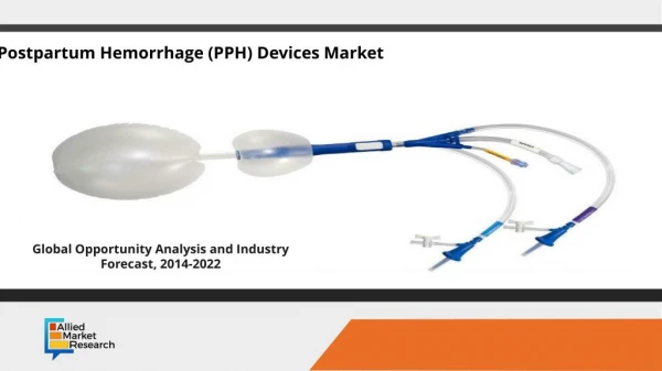
Medical Management • Bimanual uterine compression • Uterine tamponade • Packing with guaze • Can soak with thrombin • Intrauterine foley catheter • One or more bulbs, 60-80ml of saline • Bakri tamponade balloon • 300-500ml of saline
Surgical Management • Consider surgical management when uterotonic agents (± tamponade) don’t work • Uterine curettage • Exploratory laparotomy • Hypogastric artery ligation • Bilateral uterine artery ligation (O’Leary sutures) • B-Lynch technique • Hysterectomy
Other Considerations Retained Placenta • Non-adherent Retained Placenta – • Results from partial separation of a normal placenta, entrapment of the partially or completely separated placenta by an hourglass constriction ring of the uterus, mismanagement of the third stage of labor or abnormal adherence of the entire placenta or a portion of the placenta to the uterine wall. • Management : • Manual separation and removal by a primary health care provider under light anesthesia to facilitate exploration and removal
Adherent Retained Placenta • Thought to be a result from zygotic implantation in an area of defective myometrium so that there is no zone of separation between the placenta and the decidua. • Attempts to remove the placenta in the usual manner is unsuccessful, laceration or perforation of the uterine wall may result putting woman in a greater risk of PPH and infection. • Degrees of attachment: • Placenta Accreta – slight penetration of myometrium by placental trophoblast. • Placenta Increta – deep penetration of myometrium by placenta • Placenta Percreta – perforation of the uterus by the placenta • Management – • Blood component replacement therapy • Hysterectomy
Other Considerations UTERINE INVERTION • Uterus pushed “inside out”, fundus at the introitus • A rare complication 1/ 20.000 delveries • Commonly occur due to mismanagement of third stage of labour (controlled cord traction is applied when the uterus is not contract, or excessive fundal pressure) , uterine atony and uterine anomalies. –First Degree- (Incomplete)-inverted fundus reached the external os. – Second Degree- (Complete)-whole body of the uterus is inverted and protudes into the vagina – Third Degree- prolapse of inverted uterus, cervix and vagina outside the vulva
Consequences – Severe shock - anuria and renal failure – Sepsis – Chronic inversion – Uterus strangulate and slough off • Uterine inversion • If occurs prior to placental delivery, do Not remove the placenta • Replace fundus with firm pressure upwards • Uterine relaxation may be required • Terbutaline, nitroglycerine, anesthesia • Consider activation of massive transfusion protocol
Review • Stay Calm! • Tone, Tissue, Trauma, Thrombin • Postpartum hemorrhage is a symptom, not a diagnosis – find a diagnosis
Late Postpartum Hemorrhage • Blood loss of more than 500 ml. later than 24 hrs after delivery. Sometimes not occuring until 5-15 days after delivery • Clinical signs : crampy abdominal pain ,delayed uterine involution ,and signs of infection • Causes : • Sub-involution – delayed return of uterus to its pre-pregnant size and consistency. • Retained placental fragments • Predisposing Factors : • Attempts to deliver placenta before separation • Manual removal of the placenta • Placenta accreta • Uterine infection Management : Oxytocin, Methylergometrine or Prostaglandins Curettage Broadspectrum antibiotics if there is infection ( fever, uterine tenderness, foul smelling lochia )
Postpartum Infections • Puerperal Infection – • Term used to describe bacterial infections after childbirth • A fever of 38 C ( 100.4 F ) or higher after the first 24 hrs. after childbirth occuring on at least 3 of the first 10 days after the first 24 hrs. • During the first 24 hrs. a slight elevation may occur because of dehydration or the exertion of labor • Organisms can move from the vagina, cervix, uterus and out of the fallopian tube to infect the ovaries and the peritoneal cavity. Blood vessels or lymphatics can carry infection to the rest of the body.- Septicemia • Causative organisms can be Staphylococcus aureus, gonococci, coliform bacteria, and rarely by Clostridia
Risk or Predisposing Factors: • Cesarean birth increases the risk 5x because of trauma to tissues • Prolonged labor • Colonization of the vagina with pathogenic organisms • History of previous infections ( UTI, mastitis thrombophlebitis) • Trauma • Prolonged rupture of membranes • Catheterization • Excessive number of vaginal examinations • Retained placental fragments • Hemorrhage • Poor general health( fatigue, anemia, frequent minor illness) • Poor nutrition ( < PRO, Vitamin C ) • Poor hygiene • Medical conditions such as DM • Low socioeconomic status
Signs and Symptoms of Postpartum Infection • Fever and chills • Pain and redness of wounds • Purulent wound drainage or wound edges not approximating • Tachycardia • Uterine Subinvolution • Abnormal duration of lochia or foul odor • Elevated white blood cell count • Frequency or urgency of urination, dysuria or hematuria • Suprapubic pain • Localized area of warmth, redness or tenderness in the breast • Body aches, general malaise
Endometritis – • Infection of the uterus with pelvic cellulitis involving the decidua, myometrium, and parametrial tissues • Caused by organisms that are normal inhabitants of the vagina and cervix ( E. coli, bacteroids, staphylococcus, anaerobic non-hemolytic streptococcus • Signs and Symptoms : • Occurs during the first 2-7 days • Fever, chills, malaise,anorexia, abdominal pain and cramping, uterine tenderness,purulent foul smelling lochia, tachycardia, subinvolution
Management : • Broad spectrum antibiotics – IV Ampicillin, Cephalosporins, Gentamycin, Clindamycin • Antipyretics and Oxytocics – which increases drainage of lochia and involution • Complications : • Salphingitis • Oophoritis • Peritonitis • Pelvic Thrombophlebitis
Thrombophlebitis • Involves the saphenous veins and confined to the lower leg The great saphenous vein (GSV, alternately "long saphenous vein") is a large, subcutaneous, superficial vein of the leg. It is the longest vein in the body, running along the length of the lower limb. • Can be prevented by early ambulation after childbirth. • Ambulation prevents stasis of blood in the legs and decrease likelihood of thrombus formation. • Thrombus – is a collection of blood factors, platelet, and fibrin on a vessel wall. Its formation is associated with inflammatory process in the vessel wall ( Thrombophlebitis)
3 Major Causes of Thrombosis : • Venous stasis – occurs from compression of the large vessels of the pelvis and legs by the enlarging uterus • Woman stands for prolonged periods of time • Prolonged time in stirrups for delivery and repair of episiotomy Hypercoagulation - Levels of most coagulation factors are increased and fibrinolytic symptoms is suppressed which hinders clot disintegration Blood Vessel Injury - specifically to the intima of the blood vessel
Factors that increases the risk of Thrombosis : • Inactivity • Obesity • Cesarean birth • Smoking • History of previous thrombosis • Varicose veins • Diabetes Mellitus • Prolonged time in stirrups • Maternal age older than 35 years • Parity greater than 3
Clinical Manifestations : • Pain and tenderness in the lower extremity • Physical examination may revealwarmth, redness and an enlarged hardened vein over the site of the thrombosis (positive homan’s sign) • Deep vein thrombosis is more common in pregnancy and is characterized by unilateral leg pain, calf tenderness, and swelling Management : - Analgesia ( NSAID ) - Rest with elevation of the affected leg - Use of elastic stockings - Local application of heat - Anticoagulant, Antibiotics, Bedrest, Analgesia, use of elastic stockings
Peritonitis- • Inflammation of the membrane lining the walls of the abdominal and pelvic cavities and may lead to pelvic abscess.
Subinvolution of the Uterus – - refers to a slower than expected return of the uterus to its pre-pregnancy size after childbirth. - Uterus descends at the rate of 1 cm. or 1 fingerbreadth per day. By 2 weeks it should not be palpable above the symphysis pubis.
Causes of Subinvolution of the Uterus • Retained placental fragments • Pelvic infection • Signs : • Prolonged lochial discharge • Irregular or excessive uterine bleeding • Pelvic pain or feelings of heaviness • Backache, fatigue and persistent malaise • On bimanual exam, uterus feels larger and softer than normal during puerperium • Treatment : • Methylergonovine Maleate (Methergine) 0.2 mg every 3-4 hrs for 24-48 hrs. • Antimicrobial therapy
Mastitis • An infection of the lactating breast occuring during the 2nd or 3rd wk. after birth although it may develop at anytime during breastfeeding. • Common in mothers nursing for the first time and affects only one breast. • Causes : • Staphylococcus aureus or E. coli- enters thru cracks or fissures in the nipple • Engorgement and stasis of milk • Constriction of breast from too tight brassiere – may interfere with emptying of milk ducts • Fatigued and stressed out mothers
Symptoms : • Flu like with fatigue and aching muscles • Fever of 38.4C or higher • Chills, malaise and headache • Localized area of redness and inflammation • Purulent discharge – rare Management : - Antibiotic therapy - Decompression of the breast by breastfeeding or pump - Application of heat or ice packs, breast support - Analgesics
Puerperal Infections: Perineal and Cesarean wounds • Perineal wound • Assessment • Cesarean wound • Assessment • Intervention • Sutures removed • Drain purulent material • Antibiotics • Analgesics • Warm compress or sitz baths