Download
1 / 12
160 likes | 881 Views
Care Of A Patient with Brain Cancer After A Craniotomy. Brittani Allen St. Cloud State University SCH Ortho/Neuro Summer Nurse Intern 2009. Brain Facts. Primary brain tumors compose a heterogeneous group of neoplasms that

E N D
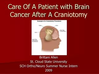
Care Of A Patient with Brain Cancer After A Craniotomy Brittani Allen St. Cloud State University SCH Ortho/Neuro Summer Nurse Intern 2009
Brain Facts Primary brain tumors compose a heterogeneous group of neoplasms that vary widely by site of origin, morphologic features, growth potential, extent of potential invasiveness and tendency for progression, and recurrence and treatment response. Types: Benign brain tumor- slow-growing cells, distinct borders, rarely spreads, may be life threatening because of its location in the brain, often needs only a craniotomy resection Malignant brain tumor- fast-growing cells, invasive of surrounding tissues, often spreads to other places in the CNS, very life-threatening, requires many therapy treatments like a craniotomy, radiation & chemotherapy Anatomical Locations: Brain, cranial nerves spinal cord, meninges, ventricle, cerebellum, pituitary, pineal, nasal cavity, other CNS, Classifications: • Tumors of neuroepithelial tissue (i.e., astrocytic tumors, oligodendroglial tumors, ependymal tumors, glioblastoma multiforme tumor) • Tumors of the meninges (i.e., meningioma) • Tumors of cranial and spinal nerves (i.e., schwannoma, neurofibroma) • Hematopoietic neoplasms (i.e., malignant lymphoma) • Germ cell tumors (i.e., teratoma) • Cysts and tumors such as lesions (i.e., Rathke cleft cyst, epidermoid cyst) • Tumors of the sellar region (i.e., pituitary adenoma) • Local extensions from regional tumors (i.e., paraganglioma, chordoma) • Metastatic tumors
Craniotomies for Brain Cancer Patients • Craniotomy with maximal surgical excision of a brain tumor provides the best treatment for prolongs survival and improving neurological status of patients with brain tumors (Sawaya, 1998). • Craniotomy for surgical resection of a brain tumor may not be curative in some cases. However, it does offer more accurate diagnosis than needle biopsy, improvement in symptoms with decreased ICP and theoretically an increased response to other treatments such as chemotherapy and radiation. • Postoperative craniotomy complications can often lead to permanent neurologic injury if gone unrecognized. Prompt recognition of neurological decline and timely diagnosis and intervention by the multidisciplinary team improves patient outcomes and subsequent quality of life.
Post-op Craniotomy Complications • Reversal agents for anesthetic complications • Needed if a pre-op LOC is not obtained • Interventions: Narcan, Flumazenal, anticholinesterase agents, anticholinergic agents • Respiratory complications • Decrease LOC, inability to protect airway • Development of edema around the brain stem • Interventions: neutral neck position, SpO2>94%, IS, ABGs, Swallow Eval • Cardiovascular complications • Hypovolemic shock: tachycardia, decreased BPs, shallow fast respirations, cool, pale skin, decreased UOP • Interventions: Fluid resuscitation, isotonic & hypertonic saline • Blood pressure: • Hypertension: may cause hemorrhage • Hypotension: may cause hypoperfusion and ischemia in surrounding brain • Cardiac arrhythmias: based on hx and increased sympathetic discharge • Interventions: obtain cardiac enzymes, check K+ & Mg+, aggressively treat MI
GI complications- gastric stress ulceration & hemorrhage • GI irritation caused by some neurological drugs (Decadron, Phenytoin) • Interventions: Antacids, H2 blockers, Sucralfate, Proton pump inhibitors. Hgb, Hct and stools should be monitored closely • Endocrine complications • Diabetes insipidus- caused by the disturbance of the pituitary gland. Due to the secretioon of an insufficient amount of ADH. Often self limiting. • Look for: polydipsia, low UOP, Na+ levels > 145 mEq/L • Interventions: replace excess UOP with IV fluids or oral fluids, Desmopresin acetate, Aqueous vasopression • Syndrome of inappropriate ADH (SIADH) • Look for: high levels of ADH, continually reabsorbed H2) from the kidney tubules, Na+ < 135 mEq/L, low UOP, increased weight • Interventions: Fluid restriction < 1000ml, replace Na+ with hypertonic saline
Infection complications • Meningitis- inflammation of the protective barrier over the brain and spinal cord • Look for: fever, headache, malaise, photophobia, hemiparesis, altered mental status, seizures, petichial rash • Interventions: pathogen-specific broad spectrum antibiotics, corticosteroid therapy, surgical intervention • Brain abscess- puss that may be encapsulated in the brain tissue usually related to infection of another body part • Look for: headache, nausea & vomiting, altered LOC, focal neurological deficits, seizures, fever, purulent discharge • Interventions: antibiotics, surgeries such as craniotomy or Burr hole aspiration, insertion of drains • Wound infection- usually caused by staphylococcal organisms • Look for: redness and drainage from wound, foul odor from wound, elevated WBC, fever, warmth and tenderness at site • Interventions: antibiotics, craniotomy for surgical irrigation & debridement, prophylactic antibiotics pre-op, intra-op & post-op • Hematological complications • DVTs- deep vein thrombosis. Incidence- 1.6%-4.0%. Causes pulmonary embolism • Look for: redness, tenderness, warmth, swelling, positive Homan’s sign • Preventative interventions: PROM, early ambulation, sequentials & TEDS, low-dose anticoagulants (Lovenox, heparin, Coumadin) • Interventions: bedrest with elevated affected extremity, heparin drip (PTT 1.5-2), vena cava filter
Neurologic complications • Hemorrhage- bleeding into the subdural, epidural, intraparenchymal, or intraventricular space • Look for: sudden hemiplegia, depressed LOC, signs and symptoms of ICP • Interventions: obtain coagulopathy labs and correct abnormalities, immediately intervene to prevent irreversible cerebral damage and death • Increase intracranial pressure- caused by hemorrhage, diffused edema, surgical trauma, hydrocephalus, retraction on brain, interference with venous drainage, cerebral infarct • Look for: decreased LOC, headache, papillary abnormalities, visual disturbances, sixth nerve palsy, hemiparesis, Cushing’s response (hypertension, respiratory irregularity, bradycardia) • Interventions: place external ventricular drainage catheter with drainage, maintain cerebral perfusion, elevate HOB to 30-45 degrees, Mannitol, hypertonic saline, hypothermia, hyperventilation
Hydrocephalus • Look for: mental status changes with lethargy and confusion, generalized weakness • Interventions: placement of ventriculostomy to drain CSF, surgical shunting • Seizures- may be generalized convulsions or focal seizure activity • Look for: hx of seizures, 40% of seizures occur with tumors in the frontal, temporal, and parietal lobes, 14% of seizures occur with tumors in the occipital lobe • Interventions: order CT and basic laboratory profile after seizure activity, obtain EEG, monitor anticonvulsant level frequently • CSF Leak • Look for: rhinorrhea or otorrhea with clear fluid, may not be at craniotomy site, salty-sweet taste in mouth • Interventions: bedrest, HOB > 30 degrees, avoid straining, Diamox, lumbar drain, surgical exploration
Common Post-op Interventions Neurological: • Head dressing & incision care- • Monitor for drainage • Change as needed, usually removed after 24 hours • Monitor incision for signs of infection • Keep staples or stitches dry • Drains- • Monitor amounts of drainage • Maintain patency • Know location of drain • Post-op imaging • Views changes from new tumor, residual tumor, or radiation effects • CT images postoperative blood or edema • MRI is baseline scan before treatment with radiation or chemotherapy Cardiovascular: • Monitor cardiac rate & rhythm • Monitor BPs Respiratory: • Prevent atelectasis & pneumonia • Maintain SaO2>94% • Encourage incentive spirometry q 2 hours at least Nutrition: • Begin with clear liquids, advance as tolerated • Swallow evaluation • Assess for nausea and vomiting, administer antiemetics as ordered.
Blood Glucose: • Hyperglycemia disrupts the blood-brain-barrier and increases edema • Steroids increase blood glucose levels • Monitor blood glucose levels before meals, at bedtime and as needed, administer hypoglycemic as needed IV Fluids: • Titrate IV fluids down once the patient is taking adequate food and liquids to prevent fluid overload & potential edema • Hypertonic solutions are used with patients with fluid restrictions and edemas • Do not use fluid with dextrose Activity: • Early ambulation is important to prevent pneumonia, atelectesis, DVTs, and deconditioning. • Physical therapy and occupational therapy consults are strongly recommended Pain: • Assess pain every 4 hours using appropriate pain scale • Administer ordered pain medications as needed • Reassess pain up to 1 hour after giving pain medication • Suggest alternative pain relief therapies (deep breathing, music, ice, darken room)
Reference American Association of Neuroscience Nurses. (2006). Guide to the Care of the Patient with Craniotomy Post–Brain Tumor Resection. Glenview, IL: Author.
Thank You! Roxanne Reining Jenni Polkhamp Ortho/Neuro Nursing Staff St. Cloud Hospital