Download
1 / 19
190 likes | 300 Views
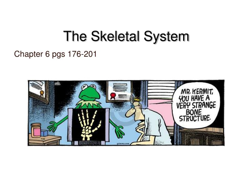
The Skeletal System. Chapter 6 pgs 176-201. Skeletal Cartilages. Basic Structure, Types, and Locations: Skeletal Cartilages- made from cartilage, surrounded by a layer of dense irregular connective tissue called the perichondrium

E N D
The Skeletal System Chapter 6 pgs 176-201
Skeletal Cartilages • Basic Structure, Types, and Locations: • Skeletal Cartilages- made from cartilage, surrounded by a layer of dense irregular connective tissue called the perichondrium • Hyaline cartilage- most abundant and includes the articular, costal, respiratory, and nasal cartilages
Skeletal Cartilages • Basic Structure, Types, and Locations: • Elastic cartilages- more flexible than hyaline and are located only in the external ear and the epiglottis of the larynx. • Fibrocartilage- located in areas that must withstand a great deal of pressure or stretch • Knee • Intervertebral discs
Skeletal Cartilages • Growth of Cartilage • Appositional growth results in outward expansion due to the production of cartilage matrix on the outside of the tissue • Interstitial growth results in expansion from within the cartilage matrix due to division of lacunae-bound chondrocytes and secretion of matrix
Classification of Bones • Two main division of the skeleton: • Axial Skeleton- Skull, vertebral column, and the rib cage • Appendicular Skeleton- Bones of the upper and lower limbs, and the girdles that attach them to the axial skeleton http://www.besthealth.com/besthealth/bodyguide/reftext/images/Axial_AppendicularSkel.jpg`
Classification of Bones: Bone Shape • Long Bone • Longer than they are wide • Have a definite shaft and two ends • Consist of all limb bones except patellas, carpals, and tarsals. • Short Bone • Somewhat cube-shaped • Include the carpals and tarsals http://www.ganfyd.org/images/4/48/Carpal.PNG http://www.arthursclipart.org/medical/skeletal/long%20bone%20structure.gif
Classification of Bones: Bone Shape • Sesamoid Bone • Special type of short bone that form in a tendon • Example: Patella (knee cap) http://www.healthhype.com/wp-content/uploads/kneecap.jpg
Classification of Bones: Bone Shape • Flat Bone • Thin, flattened, and often curved bones • Include most of the skull bones, the sternum, scapula, and ribs. • Irregular Bone • Have complicated shapes that do not fit into any other class • Vertebrae • Hip http://www.arthursclipart.org/medical/skeletal/page_04.htm
Functions of Bone • Support • Supports soft tissues and provides points of attachment for most skeletal muscles • Protection • Provides protection for the body’s vital organs • Cranial bones protect the brain • Vertebrae protect the spinal cord • Ribcage protects the heart and lungs
Functions of Bone • Movement • Muscles are attached to bones, therefore when muscles contract they cause bones to move • Mineral Storage • Stores minerals such as calcium and phosphate • Blood Cell Formation • Most blood cell formation, or hematopoiesis, occurs in the marrow cavities of certain bones
Bone Structure:Gross Anatomy • Bone Markings • Projections- grow outward from the bone surface, are sites of muscle and ligament attachment, and help form joints • Depressions and Openings- Allows blood vessels and nerves to pass and indent the bone • Types of bone markings are listed in Table 6.1 on page 179 http://classes.mst.edu/ide120/lessons/composite/materials/examples/bone_2.gif http://www.labtechindia.net/product/Biology/bl42.jpg
Bone Structure:Gross Anatomy • Bone Textures • Compact Bone- Dense outer layer that looks smooth and solid to the naked eye • Spongy Bone- Internal layer that is a honeycomb of small needle-like or flat pieces called trabeculae http://www.gla.ac.uk/ibls/US/fab/tutorial/generic/bone2.html
Bone Structure:Gross Anatomy • Epiphyses are at the ends of the bone, and consist of internal spongy bone covered by an outer layer of compact bone. • Structure of a typical long bone • Tubular bone shaft, called the diaphysis, consists of compact bone that surrounds a hollow medullary cavity, which is filled with yellow bone marrow in adults. http://www.teachpe.com/images/anatomy/bone_structure.jpg
Bone Structure:Gross Anatomy • Structure of a typical long bone • Epiphyseal line- located between the diaphysis and each epiphysis, and is a remnant of the epiphysealplate, which is a disc of hyaline cartilage that grows during childhood to lengthen the bone • Periosteum- covers the external surface of the bone • Endosteum- A connective tissue membrane that lines the internal surface of the bone
Bone Structure:Gross Anatomy Figure 6.4 page 181 • Structure of Short, Flat, and Irregular Bones • Consist of thin plates of periosteum-covered compact bone on the outside and endosteum-covered spongy bone inside
Bone Structure:Gross Anatomy • Location of Hematopoietic Tissue in Bones • Hematopoietic tissue of bones= red bone marrow • Located within the trabecular cavities of the spongy bone in flat bones and in the epiphyses of long bones. • Found in all flat bones, epiphyses, and medullary cavities of infants • In adults, distribution is restricted to flat bones and the proximal epiphyses of the humerus and femur http://www.besthealth.com/besthealth/bodyguide/reftext/images/Marrow.jpg