Download

1 / 13
130 likes | 330 Views
Overview: Shock (in adults). By: David I. Driver, MS III. Introduction. Shock is a physiologic state characterized by a significant, systemic reduction in tissue perfusion, thereby resulting in decreased tissue oxygen delivery.

E N D
Overview:Shock (in adults) By: David I. Driver, MS III
Introduction • Shock is a physiologic state characterized by a significant, systemic reduction in tissue perfusion, thereby resulting in decreased tissue oxygen delivery. • Prolonged oxygen deprivation leads to generalized cellular hypoxia and the derangement of critical biochemical processes: • Cell membrane ion pump dysfunction • Intracellular edema • Leakage of intracellular contents into the extracellular space • Inadequate regulation of intracellular pH • High Mortality • Hypovolemic – 35-40% • Cardiogenic 60-90%
Stages of Shock • The shock syndrome is characterized by a temporal continuum of physiologic stages beginning with an initial inciting event which causes a systemic circulatory disturbance; shock may subsequently progress through three stages, culminating in irreversible end-organ damage and death. • Preshock • Shock • End-organ dysfunction
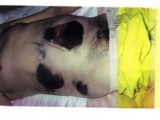
Stages of Shock II • Preshock • Preshock is also known as warm shock or compensated shock. During this stage, the body's homeostatic mechanisms rapidly compensate for diminished perfusion. Despite a 10 percent reduction in total effective blood volume, for example, a previously healthy adult may be asymptomatic. • Shock • During this stage, the regulatory mechanisms are overwhelmed and signs and symptoms of organ dysfunction appear, including tachycardia, tachypnea, metabolic acidosis, oliguria, and cool and clammy skin. • End-organ dysfunction • During this stage, progressive end-organ dysfunction leads to irreversible organ damage and death: • Urine output may decline, culminating in anuria. • Restlessness evolves into agitation, obtundation, and coma. • Acidosis further decreases cardiac output and alters cellular metabolic processes. • Multiple organ system failure proceeds to cause the demise of the patient.
Physiologic Determinants • Global tissue perfusion is determined by systemic vascular resistance (SVR) and cardiac output (CO) • SVR is governed by vessel length, blood viscosity, and the inverse of vessel diameter. • CO is the product of heart rate and stroke volume; in turn, stroke volume depends upon preload, myocardial contractility, and afterload (impedance to blood flow).
Classification of Shock • Any classification scheme simplifies the complex pathophysiology underlying the many individual causes of shock states. • Three broad types of shock states are recognized. Each type is characterized by one primary physiologic derangement: • Hypovolemic • Cardiogenic • Distributive or vasodilatory
Hypovolemic Shock • Hypovolemic shock results from decreased preload. • Two broad etiology based categories: • Hemorrhage • trauma, upper and lower gastrointestinal bleeding, ruptured aortic or ventricular aneurysm, ruptured hematoma, hemorrhagic pancreatitis, and fractures. • Fluid loss • diarrhea, vomiting, heat stroke, inadequate repletion of insensible losses, burns, and "third spacing"
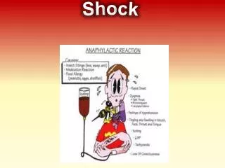
Distributive (vasodilatory) Shock • Distributive or vasodilatory shock results from a severe decrease in SVR, often associated with an increased cardiac output. • Septic shock • Activation of the systemic inflammatory response • Toxic shock syndrome • Anaphylaxis and anaphylactoid reactions • Drug or toxin reactions, including insect bites, transfusion reactions, and heavy metal poisoning • Addisonian crisis • Myxedema coma • Neurogenic shock after a central nervous system or spinal cord injury • Some patients after acute myocardial infarction who develop cardiogenic shock accompanied by a systemic inflammatory state • Post-cardiopulmonary bypass
Cardiogenic Shock • Cardiogenic shock results from pump failure, manifested physiologically as decreased systolic function and cardiac output. • Four broad categories: • Myopathic • Arrhythmic • Mechanical • Extracardiac (obstructive)
Common Features of Shock • The clinical presentation of shock varies both with the type and the cause, but several features are very common. Assessment for each of the following five major features should be done immediately in any patient suspected of developing shock. • Hypotension • Cool, clammy skin • Oligouria • Change in mental status • Metabolic acidosis
Initial Exam • History • The patient rarely provides any history; instead, historical data are usually obtained from relatives or available medical records. The patient's general condition, recent complaints, and activities prior to presentation may hold valuable information about the primary cause of shock. • Physical Exam • The physical examination should be performed rapidly and efficiently, with efforts directed toward uncovering the most likely causes of shock. • Labs • laboratory tests helps to identify potential causes for shock and early signs of organ failure
Pulmonary Artery Catheterization • Pulmonary artery catheterization is frequently used to provide hemodynamic measurements in shock patients, including CO, pulmonary artery wedge (pulmonary capillary wedge) pressure, and SVR. These values may help distinguish which type of shock is affecting a patient and can aid in identifying a specific diagnosis amongst an extensive differential.
References • 1. Barber, AE. Cell damage after shock. New Horiz 1996; 4:161. • 2. Kristensen, SR. Mechanisms of cell damage and enzyme release. Dan Med Bull 1994; 41:423. • 3. Rodgers, KG. Cardiovascular shock. Emerg Med Clin North Am 1995; 13:793. • 4. Bone, RC. Toward an epidemiology and natural history of SIRS (systemic inflammatory response syndrome). JAMA 1992; 268:3452. • 5. Moscucci, M, Bates, ER. Cardiogenic shock. Cardiol Clin 1995; 13:391. • 6. Hochman, JS, et al. Current spectrum of cardiogenic shock and effect of early revascularization on mortality. Circulation 1995; 91:873. • 7. Shoemaker, WC. Temporal physiologic patterns of shock and circulatory dysfunction based on early descriptions by invasive and noninvasive monitoring. New Horiz 1996; 4:300. • 8. • Abboud, FM. Pathophysiology of hypotension and shock. In: Hurst, JW (Ed), The Heart, New York, McGraw-Hill, 1982, p. 452. • 9. • Chien, S. Role of the sympathetic nervous system in hemorrhage. Physiol Rev 1967; 47:214. • 10. • Abboud, FM. Pathophysiology of hypotension and shock, In: Hurst, JW (Ed), The Heart, New York, McGraw-Hill, 1982, p. 452. • 11. Tuchschmidt, JA, Mecher, CE. Predictors of outcome from critical illness. Crit Care Clin 1994; 10:179. • 12. Casey, LC, Balk, RA, Bone, RC. Plasma cytokine and endotoxin levels correlate with survival in patients with the sepsis syndrome. Ann Intern Med 1993; 119:771. • 13. Chittock, DR, Russell, JA. Oxygen delivery and consumption during sepsis. Clin Chest Med 1996; 17:263. • 14. Hinshaw, LB. Sepsis/septic shock: Participation of the microcirculation: an abbreviated review. Crit Care Med 1996; 24:1072. • 15. Lederle, FA, et al. Ruptured abdominal aortic aneurysm: the internist as diagnostician. Am J Med 1994; 96:163. • 16. Kinch, JW, Ryan, TJ. Right ventricular infarction. N Engl J Med 1994; 330:1211. • 17. Hochman, JS. Cardiogenic shock complicating acute myocardial infarction: expanding the paradigm. Circulation 2003; 107:2998. • 18. Bouachour, G, et al. Hemodynamic changes in acute adrenal insufficiency. Intensive Care Med 1994; 20:138. • 19. Levraut, J, Ciebiera, JP, Chave, S, et al. Mild hyperlactatemia in stable septic patients is due to impaired lactate clearance rather than overproduction. Am J Respir Crit Care Med 1998; 157:1021. • 20. Mimoz, O, et al. Pulmonary artery catheterization in critically ill patients. Crit Care Med 1994; 22:573. • 21. Robin, ED. The cult of the Swan-Ganz catheter. Overuse and abuse of pulmonary flow catheters. Ann Intern Med 1985; 103:445. • 22. Connors, AF Jr, Speroff, T, Dawson, NV, et al. The effectiveness of right heart catheterization in the initial care of critically ill patients. SUPPORT Investigators. JAMA 1996; 276:889. • 23. Harvey, S, Harrison, DA, Singer, M, et al. Assessment of the clinical effectiveness of pulmonary artery catheters in management of patients in intensive care (PAC-Man): a randomised controlled trial. Lancet 2005; 366:472. • 24. Shah, MR, Hasselblad, V, Stevenson, LW, et al. Impact of the pulmonary artery catheter in critically ill patients: meta-analysis of randomized clinical trials. JAMA 2005; 294:1664.