Download
1 / 27
270 likes | 420 Views
Biological Bases of Behaviour. Lecture 12: Sensory Coding. Learning Outcomes. By the end of this lecture you should be able to: 1 . Describe the processes involved in sensory coding. 2 . Describe the components on the visual system

E N D
Learning Outcomes. • By the end of this lecture you should be able to: • 1. Describe the processes involved in sensory coding. • 2. Describe the components on the visual system • 3. Explain how coding takes place in the retina and cortex.
Principles of Sensory Coding. • When any stimulus (e.g. light) reaches a receptor (e.g. a retinal cell) the following events take place: • 1. Reception: Specialised cells called receptors absorb the physical energy. • 2. Transduction: Physical energy is converted into electrochemical energy represented by the firing pattern of different neurons. • Each receptor is specialised to absorb and transduce only one kind of energy i.e. visual receptors in the retina are not activated by sound. • The strength of the receptor potential (a bright spot of light or a dim spot of light) determines how strongly the receptors are activated.
3. Coding. • This refers to the one-to-one correspondence between some aspect of the physical stimulus and some aspect of neural activity. • A key aspect of sensory coding is that a given frequency of impulses in one neuron may mean something different than the same frequency of impulses in another neuron. • This is the law of specific nerve energies, (Müller, 1838). • This means that an action potential always conveys the same kind of information, i.e. the brain 'sees' the activity of the optic nerve but 'hears' the activity of the auditory nerve. • Flashes of light can be seen when the eye is pressed - the brain interprets any stimulation to the retinal receptors as 'light' even when there is none present.
Qualifications. • a) Cells with a spontaneous firing rate may signal one kind of stimulus by increasing their firing rate, and a different kind of stimulus by a decrease in their firing rate. • b) In some cases, information depends upon the timing of action potentials and not just their total number. E.g in the retina, neuron A firing just before neuron B may signal movement. • c) The exact meaning of an impulse in a single neuron depends upon which other neurons are active, so the activity of a visual neuron may contribute to the sensation of green or yellow depending upon the activity of other neurons.
Synaesthesia. • If it were possible to swap the auditory and optic nerves then we may 'see’ sounds and 'hear' lights. • A neurological disorder called synaesthesia may actually indicate that such rewiring is possible. • Here, a stimulus presented in one sensory modality (sound) triggers sensations in another sensory modality (vision). • A synaesthete may describe the colour of someone’s voice; taste shapes, or describe music in visual terms (Baron-Cohen et al., (1987). • Baron-Cohen et al., (1993) argued that this condition may be caused by connections between colour and hearing modules that have not died off (in normal development regions share many interconnections which are then pruned).
4. Awareness. • Most stimuli that are received, transduced and coded are then perceived. • E.g, when smelling a flower, scent molecules strike olfactory receptors in the nose (reception). • This produces a chemical reaction that depolarizes the resting potentials of the olfactory receptors, they fire (transduction) and this information is passed via the olfactory nerve to the olfactory bulb at the base of the brain (coding). • The olfactory bulb then sends connections to various parts of prefrontal cortex where smells are recognised (awareness).
Example: The Visual System. • Light enters the eye through an opening in the centre of the iris called the pupil. • The light is focused by the cornea and lens and projected onto the retina - the light sensitive cells that line the rear of the eyeball. • Light from the top left of the visual scene strikes the bottom right of the retina and vice versa so the visual image on the retina is upside down and reversed. • The centre of the retina is called the macula and this is the most sensitive part of the retina used for resolving fine detail. • The most precise region of visual analysis takes place within the macula at the fovea, a small region where receptors are tightly packed.
Anatomy of the Eye fovea iris macula pupil cornea lens Optic nerve retina Kalat (2001) p154
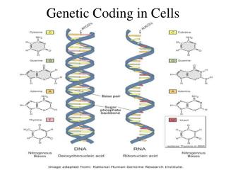
Retinal Receptors. • The photoreceptors lie on the inner surface of the retina facing away from the light source. There are two types: • Rods: Highly sensitive to dim light, and found at the periphery of the retina. They are highly sensitive to movement but have little colour coding capabilities. • Cones: Are found mainly in the fovea, are highly sensitive and used for precise vision. There are three types (red, blue and green) which are maximally responsive to these colours. They do not work well in dim light. • Rods and cones contain chemicals that release energy when struck by light (photopigments). They consist of a derivative of vitamin A called 11-cis-retinal, which is stable in the dark, but is converted to all-trans-retinal by light.
Other Retinal Cells. • Rods and cones connect to the bipolar cells, which receive support from the horizontal cells and the amacrine cells. In turn the bipolar cells induce action potentials in the ganglion cells, of which there are two types: • Magnocellular (M) cells: These are large and are found mainly in the periphery of the retina, and so receive their input mainly from rods. They are thus sensitive to light and movement, but not to colour. • Parvocellular (P) cells: These are smaller, and are found mainly in the fovea. They receive their input mainly from cones and so are sensitive to colour and fine detail. • The axons of both M and P cells form the optic nerve, which leaves the retina at the optic disc or blind spot where there are no receptors.
Retinal Cells. Carlson (2001) p165
The Retina. Blood vessels Blind spot Horizontal cell Optic nerve Amacrine cell Ganglion cells Bipolar cells Rods and cones Kalat (2001) p155
Coding in the Retina. • The human retina contains around 120 million rods and 6 million cones but we do not individually process 126 million bits of information. • Each ganglion cell has a receptive field whose size and sensitivity depends upon how many rods or cones converge upon it. • In the macula only a few cones converge upon each ganglion cell so visual acuity is enhanced. • In the periphery, many rods converge upon each ganglion cell so sensitivity is reduced. • The receptive fields of the ganglion cells converge to form the receptive fields at the next neural level and so on.
The Ganglion Cells. • In the 1930's Hartline discovered that the retina contains 3 types of ganglion cells: • On cells: these respond when a light strikes the retina. • Off cells: these respond when the light is removed. • On/off cells: these respond briefly when the light is on and also again briefly when the light is switched off. • In the 1950's Kuffler recorded the activity of retinal ganglion cells and discovered that their receptive fields are in the form of a central region surrounded by a concentric circle. • Stimulation of the centre or the surround had different effects depending upon the type of the cell:
Activity of Ganglion Cells. • On-centre: Light falling on the centre of the receptive field stimulates the cell, but light falling in the surround inhibits the cell. • Off-centre: Light falling on the surround stimulates the cell but light falling on the centre inhibits the cell. • At rest all ganglion cells fire spontaneously at a low rate, but when light falls across their receptive fields 'on cells' signal an increase in illumination, 'off cells' signal a decrease. • Receptive fields overlap which ensures that a small spot of light will excite or inhibit many ganglion cells - this is how we determine shapes. • It also ensures that the visual system is primed to perceive edges - even where none actually exist.
On/Off Centre Cells. Carlson (2001) p171
On/Off Cells in Action. Look at the centre of the grid - can you see black blobs at the intersections? Kalat (2001) p167
Perception of Edges. The ganglion cells in the retina enhance the contrast between edges, the squares appear to be very different in tone but actually are not Carlson (2001) p171
Visual Pathways. • The optic nerves from both eyes meet at the optic chiasm. • Here fibres from both visual fields in each eye cross over to be represented in opposite hemispheres. • All the P ganglion cell axons and some of the M ganglion cell axons project to the lateral geniculate nucleus (LGN) a part of the thalamus specialised for visual perception. • The LGN then sends projections to the visual (striate) cortex. • This pathway is called the geniculostriate system. • Remaining M ganglion cell axons connect to the superior colliculus (of the tectum) then to part of the thalamus called the pulvinar, and then on to visual regions in temporal and parietal cortices. • This pathway is called the tectopulvinar system.
The Visual Pathways Striate cortex Superior colliculus Lateral geniculate nucleus Optic chiasm Retina Optic nerve Kalat (2001) p165
Visual Cortex. • Most information from the LGN goes first to primary visual cortex (V1), which is responsible for the basic elements of processing. • This region then sends information to: • Secondary visual cortex (V2). • Visual association areas (V3-V5). • Temporal and parietal cortices. V3 V1 V2 V4 V5 Kolb & Whishaw (2001) p293
Coding in Visual Cortex. • Individual cells in V1 receive input from many ganglion cells and so their receptive fields are large, they do not thus respond just to 'light on' or 'light off', but respond to bars of light oriented in a particular direction - they are thus orientation or feature detectors. • In the 1950's Hubel and Wiesel discovered three types of cell: • Simple cells: these responded to the presence of a bar of light at a particular orientation and position. • Complex cells: these responded to bars of particular orientations moving across the retina. • Hypercomplex cells: these also responded to moving bars but also had a strong inhibitory region at their end.
Coding in Visual Cortex Continued. • The 3 types of cell have different tunings, some respond maximally to a thin bar of light, others to a thicker bar etc. • The cells are organised into columns within which neurons respond to similar features. • Neurons in adjacent columns respond to slightly different features. • Information from each eye is split between adjacent columns referred to as ocular dominance columns. Simple cell Complex cell Hypercomplex cell Carlson (2001) p171
Coding in Other Cortical Regions. • Cells in striate cortex only provide basic visual analysis - (shape, orientation), perception (awareness) take place in additional regions specialised for different processing. • Mishkin et al., (1983) described two distinct visual pathways which emerge from primary visual cortex: • Dorsal stream (‘where’ pathway): This travels to the parietal lobes and is important for perceiving where an object is located. Damage to it impairs the ability to locate objects even though they are perceived. • Ventral stream (‘What pathway’): This travels to the temporal lobes and is important for perceiving what an object is. Damage to it impairs visual object recognition (agnosia); e.g. a patient may be able to describe an object but not to be able to recognise what it is.
‘What’ and ‘Where’ Pathways. Parietal lobe Visual cortex Temporal lobe Kolb & Whishaw (2001) p290
References and Bibliography • Baron-Cohen, S., Wyke, M., & Binnie, C. (1987). Hearing words and seeing colours: an experimental investigation of a case of synaesthesia. Perception, 16: 761 - 767. • Carlson, N.R. (2001). Physiology of Behaviour. • Baron-Cohen, S., Harrison, J., Goldstein, L.H., & Wyke, M. (1993). Coloured speech perception: is synaesthesia what happens when modularity breaks down? Perception, 22: 419 - 426. • Kalat, J.W. (1995). Biological Psychology. • Kolb, B., & Whishaw, I.Q. (2001). Fundamentals of Human Neuropsychology. • Mishkin, M., Ungerleider, L.G., & Macko, K.A. (1983). Object vision and spatial vision: two cortical pathways. Trends in Neurosciences, 6: 414-417.