Download
1 / 48
510 likes | 639 Views
Explore cell-to-cell interactions through surface markers, junctions, neuro signaling, and GPCR mechanisms in this detailed overview.

E N D
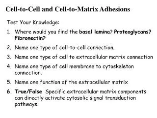
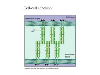
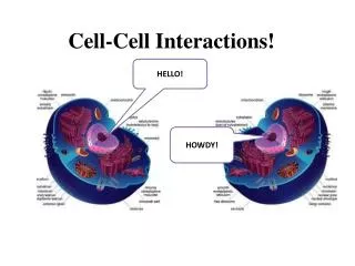
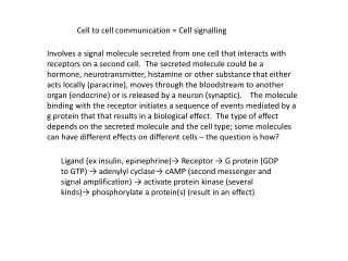
35 Cell-to-Cell Interactions Cells can identify each other by cell surface markers. -glycolipids are commonly used as tissue-specific markers -major histocompatibility complex (MHC) proteins are used by cells to distinguish “self” from “non-self”
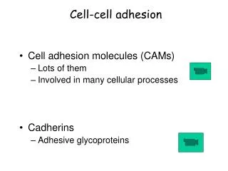
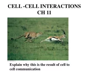
Cell-to-Cell Interactions Cells within a tissue are connected to each other by cell junctions 1. tight junctions – create sheets of cells 2. anchoring junctions – connect the cytoskeletons of adjacent cells 3. communicating junctions – permit small molecules to pass between cells a. gap junctions – in animal cells b. plasmodesmata– in plant cells
Acetylcholine: common neurotransmitter opens ligand-gated Na+ channels on muscle cell and some nerve cells
Signal transmitted to muscle cell across a synapse Musclecell a. a. Depolarization opens voltage-gated Ca+2 channels Musclecell b. Ca+2 rushes in; Vesicles fuse with membrane b. c. Neurotransmitter released; opens ligand-gated Na+ channels on muscle cell Depolarizes muscle cell c. Signal: electrical to chemical to electrical
GPCRs that Regulate Ion Channels: Muscarinic Acetylcholine Receptor The neurotransmitter, acetylcholine (ACH) binds to two types of receptors known as the nicotinicand muscarinicacetylcholine receptors. The nicotinic receptor is itself a ligand-gated ion channel that opens on ACH binding. This receptor is located in the neuromuscular junctions of striated muscle. The muscarinic ACH receptor, is a GPCR found in cardiac muscle cells that is coupled to an inhibitory G protein
The binding of ACH to this receptor triggers dissociation of Gai-GTP from Gßg, which in this case, directly binds to and opens a K+ channel. The movement of K+ down its concentration gradient to the outside of the cell, increases the positive charge outside the membrane, hyperpolarizing the cell. This results in the slowing of heart rate.
Structure of GPCRs G protein-coupled receptors (GPCRs) are the most numerous class of receptors in most eukaryotes. Receptor activation by ligand binding activates an associated trimeric G protein, which in turn interacts with downstream signal transduction proteins. All GPCRs are integral membrane proteins that have a common 7 transmembrane segment structure (Fig.( The hormone/ligand binding domain is formed by amino acids located on the external side of the membrane and/or membrane interior (Fig.). GPCRs interact with G proteins via amino acids in the C3 and C4 cytoplasmic regions.
G Protein Activation of Effectors The trimeric G protein cycle of activity in hormone-stimulated GPCR regulation of effector proteins is summarized in (next slide). Initially, the G protein complex is a chain to the inner leaflet of the cytoplasmic membrane via lipid anchors attached to the Ga and Gg subunits. The trimeric GDP-bound form of the G protein is inactive in signaling. The binding of a hormone to the GPCR triggers a conformational change in the receptor (Step 1) which promotes its binding to the trimeric G protein (Step 2). Binding to the activated GPCR triggers the dissociation of GDP (Step 3). Subsequent binding of GTP to the Ga subunit activates it, and causes its dissociation from the receptor and the Gßg complex (Step 4). Ga-GTP then binds to the effector protein regulating its activity. The hormone eventually dissociates from the receptor (Step 5). Over time (often less than 1 min), GTP is hydrolyzed to GDP and Ga becomes inactive. It then dissociates from the effector and recombines with Gßg (Step 6). A hormone-bound GPCR activates multiple G proteins, until the hormone dissociates. Proteins known as regulators of G protein signaling (RGS) accelerate GTP hydrolysis by Ga decreasing the time-period during which Ga is active (not shown).
Trimeric G Proteins & Their Effectors There are 21 different Ga proteins encoded in the human genome. The G proteins containing these subunits are activated by different GPCRs and regulate a variety of different effector proteins . The most common effectors synthesize second messengers such as cAMP, IP3, DAG, and cGMP. In the case of cAMP, a stimulatory Gs subunit activates adenylyl cyclase and cAMP production, whereas an inhibitory Gi subunit inhibits adenylyl cyclase and cAMP production.
GPCRs That Bind Epinephrine Epinephrine is a hormone that signals the "fight-or-flight" response. It elevates heart rate, dilates the airway, and mobilizes carbohydrate and lipid stores of energy in liver and adipose tissue. In the heart, liver, and adipose tissue, these effects are mediated via binding to ß1- & ß2-adrenergic GPCRs. Both ß-adrenergic GPCRs signal via Gas, which activates adenylyl cyclase and raises intracellular [cAMP]. The a2-adrenergic GPCR signals via Gai, decreasing adenylyl cyclase activity and intracellular [cAMP]. The a1-adrenergic GPCR is coupled to Gaq, which activates phospholipase C (PLC) and signaling via the IP3/DAG pathway . a1-adrenergic GPCRs are present in the liver and blood vessels in peripheral organs. Binding to a1-adrenergic GPCRs stimulates glycogen breakdown in the liver, while blood flow to peripheral organs is decreased.
GPCRs that Regulate Adenylyl Cyclase Adenylyl cyclase is an effector enzyme that synthesizes cAMP. Ga-GTP subunits bind to the catalytic domains of the cyclase, regulating their activity. Gas-GTPactivates the catalytic domains, whereas Gai-GTPinhibits them. A given cell type can express multiple types of GPCRs that all couple to adenylyl cyclase. The net activity of adenylyl cyclase thus depends on the combined level of G protein signaling via the multiple GPCRs. In liver, GPCRs for epinephrine and glucagon both activate the cyclase. In adipose tissue , epinephrine, glucagon, and ACTH activate the cyclase via Gas-GTP, while PGE1 and adenosine inactivate the cyclase via Gai-GTP.
Activation of Gene Transcription by GPCR Signaling GPCRs regulate gene transcription by cAMP and PKA signaling. As shown in the figure, cAMP-released PKA catalytic domains enter the nucleus and phosphorylate the CREB (CRE-binding) protein, which binds to CRE (cAMP-response element) sequences upstream of cAMP-regulated genes. Only phosphorylated p-CREB has DNA binding activity. p-CREB interacts with other TFs to help assemble the RNA Pol II transcription machinery at these promoters. In liver, glucagon signaling via this pathway activates transcription of genes needed for gluconeogenesis.
Down-regulation of GPCR/cAMP/PKA Signaling • A number of events contribute to the termination of signaling by a GPCR. These include: • dissociation of the hormone from the receptor, • hydrolysis of GTP by Ga • hydrolysis of cAMP via cAMPphosphodiesterase, • phosphorylation and “desensitization” of receptors by kinases such as PKA and ß-adrenergic receptor kinase (BARK). • In addition, GPCRs can be removed from the membrane by vesicular uptake.
Biological functions mediated by 7TM receptors Smell Taste Vision Neurotransmission Hormone secretion Exocytosis Control of blood pressure Embryogenesis Cell growth and differentiation Development Viral infection Carcinogenesis
G-protein activation “molecular switch” inactive • (b) Ligand binds • G-protein associates • (c) GDP-GTP exchange • -Subunit dissociates Active G-Protein-GTP -> allosteric modulator of target effector enzyme active
All G-proteins – similar structure/activation • There are TWO broad subclasses of • trimeric G-protein-activated signal • transduction pathways: • depends on theirtarget effector enzymes • A. adenylyl cyclase • B. phospholipase C
First messenger (signal molecule such as epinephrine) Adenylyl cyclase G protein GTP G-protein-linked receptor ATP cAMP Protein kinase A Cellular responses An activated Ga-protein-GTP • Can trigger the formation of cAMP, which then acts as a second messenger in cellular pathways
G-protein-GTP activation of Effector Enzyme adenylylcyclase produces the 2nd messenger cAMP Activated G-protein
Adenylyl Cyclase & Protein Kinase A Adenylyl cyclase is an integral membrane protein that contains 12 transmembrane segments (Fig.). It also has 2 cytoplasmic domains that together form the catalytic site for synthesis of cAMP from ATP. One of the primary targets of cAMP is a regulatory kinase called protein kinase A (PKA), or cAMP-dependent protein kinase.
PKA exists in two different states inside cells (Fig.). In the absence of cAMP, the enzyme forms a inactive tetrameric complex in which 2 PKA catalytic subunits are non-covalently associated with 2 regulatory subunits. When cAMP concentration rises, cAMP binds to the regulatory subunits which undergo a conformational change, releasing the active catalytic subunits.
Phosphorylase kinase inactive + P active Protein Kinase A Phosphorylates downstream target enzymes Breaks down Glycogen Into Glucose
What are targets for Protein Kinase A?? cAMP regulated pathways Function target tissue signal Glycogen breakdown muscle epinephrine Glycogen breakdown liver glucagon Heart rate cardiovascular epinephrine Water reabsorption kidney antidiuretic hormone
How to shut it off? No ligand G-protein -subunit is on a timer Inherent GTPase activity Auto Shut-off
How to shut it off? cAMP-phosphodiesterase rapidly cleaves cAMP (so short lived)
How do you turn it off? kinases – phosphatases Diametrically Opposed… Remember: whether you active or inactivate by adding P depends on the specific protein
What if you can’t turn off cascade? Vibrio cholera - causes cholera Normal gut: H20, NaCl, NaHCO3 secretion controlled by hormones via Gs/cAMP signal pathways V. cholera – secretes enterotoxin, chemically modifies Gs – no GTPase activity - stays ON Severe watery diarrhea – dehydration, death
target effector enzyme is Phospholipase C PLC cleaves a membrane phospholipid (Phoshatidyl inositol) to two 2nd Messengers: Inositol-1,4,5-Trisphosphate (InsP3) & Diacylglycerol (DAG)
DAG Lipid Soluble InsP3 Water Soluble PIP2
GPCRs That Activate Phospholipase C Another common GPCR signaling pathway involves the activation of phospholipase C (PLC). This enzyme cleaves the membrane lipid, phosphatidylinositol 4,5-bisphosphate (PIP2) to the second messengers, inositol 1,4,5-trisphosphate (IP3) and diacylglycerol (DAG) (Fig.). In this case, the Go and Gq G proteins conduct the signal from the GPCR to PLC. This is the pathway used in a1-adrenergic GPCR signaling in the liver. *
IP3/DAG Signaling Elevates Cytosolic Ca2+ The steps downstream of PLC that make up the IP3/DAG signalingpathway are illustrated in the figure. IP3 diffuses from the cytoplasmic membrane to the ER where it binds to and triggers the opening of IP3-gatedCa2+channels (Steps 3 & 4). Another kinase, protein kinase C (PKC) binds to DAG in the cytoplasmic membrane and is activated (Step 6). In liver, the rise in cytoplasmic [Ca2+] activates enzymes such as glycogen phosphorylase kinase, which phosphorylates and activates glycogen phosphorylase. Glycogen phosphorylase kinase is activated by Ca2+-calmodulin. In addition, PKC phosphorylates and inactivates glycogen synthase.
DAG Activates Protein Kinase C (Starts Cascade) InsP3 Ligand for ER ligand- gated Ca++ channels Ca++ levels
Response: Protein Kinase C phosphorylates target proteins (ser & thr) cell growth regulation of ion channels cytoskeleton increases cell pH Protein secretion Ca++ Binds & activates calmodulin Calmodulin-binding proteins activated (kinases & phosphatases)
Signal Trans. Components: GTPase Switches GTPase switch protein also play important roles in intracellular signal transduction . GTPases are active when bound to GTP and inactive when bound to GDP. The timeframe of activation depends on the GTPase activity (the timer function) of these proteins. Proteins known as guanine nucleotide-exchange factors (GEFs) promote exchange of GTP for GDP and activate GTPases. Proteins known as GTPase-activating proteins (GAPs), stimulate the rate of GTP hydrolysis to GDP and inactivate GTPases. .
We will cover two classes of GTPase switch proteins--trimeric (large) G proteins, and monomeric (small) G proteins. Trimeric G proteins interact directly with receptors, whereas small G proteins interact with receptors via adaptor proteins and GEFs.
Signal Trans. Components: 2nd Messengers While there are a large number of extracellular receptor ligands ("first messengers"), there are relatively few small molecules used in intracellular signal transduction ("second messengers"). In fact, only 6 second messengers occur in animal cells. These are cAMP, cGMP, 1,2-diacylglycerol (DAG), and inositol 1,4,5-trisphosphate (IP3) , and calcium and phosphoinositides. Second messengers are small molecules that diffuse rapidly through the cytoplasm to their protein targets. Another advantage of second messengers is that they facilitate amplification of an extracellular signal.
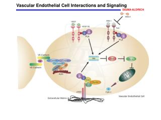
Nitric Oxide (NO)/cGMP Signaling A related signaling pathway involving phospholipase C operates in vascular endothelial cells and causes adjacent smooth muscle cells to relax in response to circulating acetylcholine (Fig.) In the NO/cGMP signaling pathway, the downstream target of Ca2+/calmodulin is nitric oxide synthase, which synthesizes the gas NO from arginine. NO diffuses into smooth muscle cells and causes relaxation by activating guanylyl cyclase and increasing [cGMP]. As a result arteries in tissues such as the heart dilate, increasing blood supply to the tissue. NO also is produced from the drug nitroglycerin which is given to heart attack patients and patients being treated for angina.
„Second messenger“ DAG, IP3 and Ca++ 10-3 M 10-7 M
Metabolism of Diacylglycerol. Diacylglycerol may be (1) phosphorylated to phosphatidate or (2) hydrolyzed to glycerol and fatty acids.
Summary • - signaling is endocrine, paracrine, synaptic, or direct cell contact • signal transduction is mediated by receptor proteins • Receptors bind primary signal (ligand) • Some amplification event occurs • Example: ligand gated ion channel opens • influx of ions triggers change in activity • (vesicle fusion in nerve end, contraction in muscle) • Example: ligand binds to 7-pass membrane receptor • catalyzes GTP exchange • to Ga-subunit of trimeric G-protein • active Ga-subunit-GTP is allosteric activator of • effector enzymes: • - ADENYLATE CYCLASE: makes cyclic AMP • - PHOSPHOLIPASE C: makes DAG and IP3 • these second messengers activate target enzymes • Trigger cascades • Must shut off cascade: removal of ligand, hydrolysis of GTP, • phosphodiesterase, protein phosphatases, Ca++ ion pumps