Download
1 / 18
180 likes | 198 Views
This project, funded by the FP7 program, focuses on developing new biomarkers for cancer detection, particularly pancreatic and prostatic cancer. The innovative dual-modality PET-US endoscopic probe offers exceptional spatial resolution of 1mm, timing resolution of 200ps, and high sensitivity for early tumor detection. The consortium overcame technical challenges such as non-conventional PET configurations and miniaturization to create cutting-edge solutions like thin crystal pixels and TOF electronic collimation. The project also addresses anatomical constraints and crystal performance optimization for superior imaging results. With an overview of the sophisticated readout architecture and data rates, the project organization spans multiple work packages involving renowned institutions to achieve its objectives of advancing image-guided interventions.

E N D
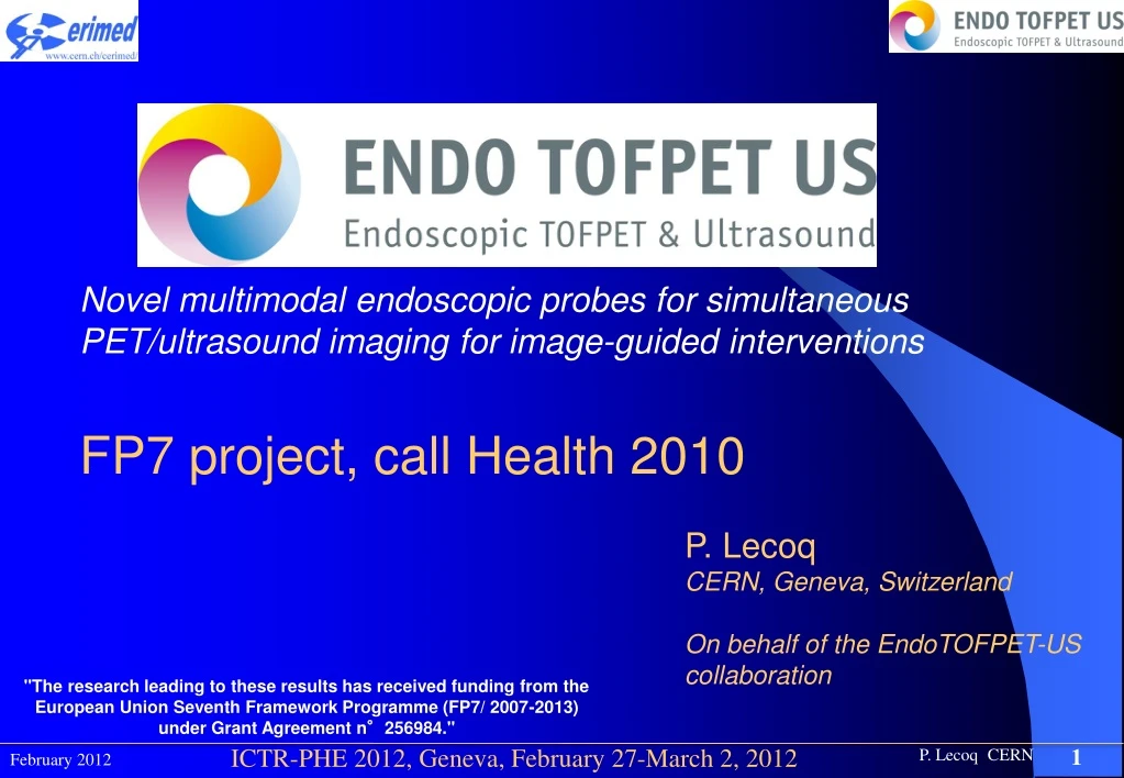
Novel multimodal endoscopic probes for simultaneous PET/ultrasound imaging for image-guided interventions FP7 project, call Health 2010 P. Lecoq CERN, Geneva, Switzerland On behalf of the EndoTOFPET-US collaboration "The research leading to these results has received funding from the European Union Seventh Framework Programme (FP7/ 2007-2013) under Grant Agreement n°256984."
EndoTOFPET-US project • Development of new biomarkers • First clinicaltargets: pancreatic/prostatic cancer • Tool: dual modality PET-US endoscopic probe • Spatial resolution: 1mm • Timing resolution: 200ps • High sensitivity to detect 1mm tumor in a few mn • Energyresolution: discriminate Compton events
Technical challenges • Non-conventional PET configuration • Assymetric: one PET head in near contact to ROI • Simulation and reconstruction problems • Endoscopic: one PET head inside the body • Miniaturization • High background from other organs (heart, bladder,…) • Variable geometry
Technical challenges • Innovative solutions • Very thin crystal pixels for the internal probe (mpulling-down crystal fibers production technology?) • for high granularity of the internal probe and <1mm spatial resolution • TOF electronic collimation with < 200ps timing resolution • for background rejection outside 3cm ROI • Digital light detection: SiPM with single SPAD readout • for single optical photon counting and ultimate timing resolution • Diffractive optics light concentrators between crystals and SiPM • for optimizing light collection • High level of electronics and mechanical integration (5mm precision) • For miniaturization • Tracking of all movables parts • for <1mm determination of their relative positions
External Plate • 20.5x20.5x1.5 cm3 • 4096 LYSO crystals 3x3x15 mm3 • 64 matrices of 64 crystals each • Hamamatsu 50mm SiPM 8x8 matrix
Endoscopic probe: crystals PET head US Probe with biopsy needle EM tracking sensor PROBE 9x18 LYSO or LSO:Ce, Ca matrix 0.75x0.75x10mm3 crystals 80mm 3M ESR gap EXTERNAL PLATE 20.5x20.5 cm2 LYSO:Ce matrix 3x3x15mm3 crystals 80mm 3M ESR gap
Crystal performance ENDOSCOPIC PROBE EXTERNAL PLATE 16 LYSO crystals matrix 0.75x0.75x10mm3 80mm 3M ESR wrapping Uniformly irradiated by a 137Cs source DRY contact to PMT 2020Q 16 LYSO crystals matrix 3x3x15mm3 80mm 3M ESR wrapping Uniformly irradiated by a 137Cs source DRY contact to PMT 2020Q
Endoscopic probe: A new concept of fully digital SiPM 30um 50um 800um 800um 14.4mm 7.2mm PET head US Probe with biopsy needle EM tracking sensor 16x26 SiPM-like or CLUSTER 48 TDCs per crystal 864 TDCs on the chip Considering also active quenching 9x18 IMAGER
Optical coupling system Collimator Detector Lenticular lens Crystal 500mm thick light concentrator glued on the crystal & photodetctor Simulations predict ayield of 68-70% (gain of ~1.7) Fill factor of the photodetector (~41%))
Overview of readout architecture DETECTOR data SPAD SPAD 1 SPAD readout & control clock, reset/sync, control 2 PCIe interface 1 Backend readout & control ASIC DAQ Server Frontend readout & control 1 16 ASIC 1 ASIC Frontend readout & control 4 16 ASIC
Event and DAQ rates • The hit rate in the external plate is expected to be 10 kHz per crystal. The total hit rate is 40 MHz. • The event rate in the internal probe is of the order of 1 MHz. • The total average data rate from the PET plate is 2 Gb/s. The available bandwidth is 3.2 Gb/s (4 x LVDS links at 800 Mb/s each) • The total average rate from the internal probe is of the order of 1 Gb/s. A total bandwidth of 1.6 Gb/s is available (2 x LVDS links at 800 Mb/s each) • Random coincidence rate is estimated at 250 kHz (assuming 6.25 ns coincidence window). The true coincidence rate is assumed to be of the same order. Therefore we assume that the total trigger rate is 0.5 MHz. • For a trigger rate of 0.5 MHz the storage rate is relatively modest (about 100 Mbyte/s). • The stored data is reduced by a factor 10 after offline filtering with a coincidence window of 600 ps.
Can we achieve 200ps timing resolution? F. Powolny et al., IEEE Trans. Nucl. Sc., Vol 58, N°3, pp597-604, June 2011 More data in: Auffray et al. Proceedings of the IEEE NSS/MIC conference, Valencia, Oct. 2011
Project Organization 4 years project WP2: CERN Crystals and optics Scintillating fibers and diffrative coupling optics WP5: DESY Detector Integration Miniaturized probe Tracking&Image fusion WP3: Delft TU Photodetectors Novel digital photodetectors WP4: LIP FE and DAQ electronics Highly integrated TOF electronics WP6: TUM Clinical requirements & preclinical and pilot clinical studies Feasibility tests on pigs, Pilot clinical tests, Impact on biomarker studies WP1: UnivMed Project Coordination
Conclusion • EndoTOFPET-US is an approved FP7 project running since 1 year • Objectives • Develop a multimodal imaging/interventional endoscopic probe based on several technological breakthroughs • To be used a a tool to develop biomarkers in priority for pancreas and prostate • Define a roadmap for the development of a new generation of multimodal endoscopic probes for different clinical applications "The research leading to these results has received funding from the European Union Seventh Framework Programme (FP7/ 2007-2013) under Grant Agreement n°256984."