Download
1 / 26
260 likes | 284 Views
Medical Microbiology Respiratory System - Pneumonia Staphylococcal Pneumonia. Dr. Sameer Naji, MB, BCh, PhD (UK) Dean Assistant Head of Basic Medical Sciences Dept. Faculty of Medicine The Hashemite University. Definition of Pneumonia.

E N D
Medical MicrobiologyRespiratory System - PneumoniaStaphylococcal Pneumonia Dr. Sameer Naji, MB, BCh, PhD (UK) Dean Assistant Head of Basic Medical Sciences Dept. Faculty of Medicine The Hashemite University
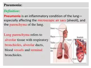
Definition of Pneumonia • An acute infection of the pulmonary parenchyma that is caused by bacteria, viruses, fungi, or parasites. It is characterized primarily by inflammation of the microscopic air sacs (alveoli). The air sacs may fill with fluid or pus (purulent material), causing cough with phlegm (sputum) or pus, fever, chills, and difficulty breathing, accompanied by an acute infiltrate on CXR or auscultatory findings consistent with pneumonia • Pneumonia can range in seriousness from mild to life-threatening. It is most serious for infants and young children, people older than age 65, and people with health problems or weakened immune systems
Classification • I. Etiology A. Bacterial Causes: Gram-positive organisms • Streptococcus • Staphylococcus Gram-negative organisms • Haemophilus influenzae • Klebsiella • Pseudomonas aeruginosa • Moraxella catarrhalis • Escherichia coli • Serratia species • Enterobacter species • Acinetobacter species
B. Atypical organisms • Mycoplasma pneumoniae • Legionella pneumophila • Chlamydia psittaci • Chlamydia pneumoniae C. Anaerobic bacterial infections • Peptostreptococcusspecies • Bacteroidesmelaninogenicus • Fusobacterium necrophorum • Bacteroidesasaccharolyticus • Porphyromonasendodontalis • Porphyromonasgingivalis
D. Viral causes • Influenza virus • Para-influenza virus • Respiratory syncytial virus • Corona virus (SARS) • MERS • Human metapneumovirus
E. Other causes • Rickettsial infections • Varicella • Rubella • Aspiration pneumonitis • Lipoid pneumonitis • Pneumocystis Jeruvici (carinii) • Cytomegalovirus • Tuberculosis • Fungal infections
II. Variants • Community-acquired pneumonia (CAP) • Nursing home–acquired pneumonia • Nosocomial pneumonia (Hospital-acquired pneumonia) when patient was hospitalized with any another diagnosis, and after 48 hours in the hospital (not earlier!) pneumonia was diagnosed, or pneumonia after artificial lung ventilation (Ventilator-associated pneumonia) • Pneumonia due to aspiration. It results from the aspiration of gastric contents in addition to aspiration of upper respiratory flora in secretions. • Pneumonia in immunocompromised host – patients with AIDS or immunodeficit of other origin. Causes of pneumonia – viruses, fungi of saprofites (E. coli etc.)
Community acquired Streptococcus pneumoniae Mycoplasma pneumoniae Influenza virus A Haemophilus influenzae Legionella pneumophila Staphylococcus aureus Coxiellaburneti Chlamydia psittaci Hospital acquired Gram-negative bacilli Staphylococcus aureus (+MRSA) Streptococcus pneumoniae Legionella pneumophila Haemophilus influenzae Pseudomonas spp Acinetobacter species Klebsiellapneumoniae Serratiamarcescens Immunocompromised patients Pneumocystis carinii Cytomegalovirus Mycobacterium avium-intracellulare Mycobacterium tuberculosis Streptococcus pneumoniae Haemophilus influenzae Legionella pneumophila Actinomycesisraelii Aspergillus fumigatus Nocardiaasteroides Pneumonia: infecting organisms in approximate descending order of frequency
Risk Factors for pneumonia • age • alcoholism • smoking • asthma • immunosuppression • institutionalization • COPD • PVD • Dementia
Risk Factors for Mortality • age • bacteremia (for S. pneumoniae) • extent of radiographic changes • degree of immunosuppression • amount of alcohol
Clinical manifestations • Shaking chills • Rapidly rising fever ( 39.5 to 40.5 degree) • Stabbing chest pain aggravated by respiration and coughing • Tachypnea, nasal flaring • Patient is very ill and lies on the affected side to decrease pain • Use of accessory muscles of respiration e.g. abdomen and intercostals muscles • Cough with purulent, blood tinged, rusty sputum • Shortness of breath • Flushed cheeks • Loss of appetite, low energy, and fatigue • Cyanosed lips and nail beds • Diarrhea (Legionella) • URI, sinusitis (Mycoplasma)
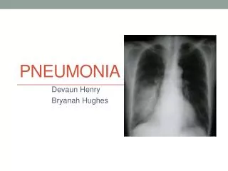
Diagnostic Tests • History taking • Physical examination • Chest x-ray • Blood test • Gram Stain and Sputum culture • Blood Culture • Antigen Tests
Gram's Stain and Culture of Sputum • a sputum sample must have >25 neutrophils and <10 squamous epithelial cells per low-power field • sensitivity / specificity of the sputum Gram's stain and culture are highly variable • in cases of proven bacteremicpneumococci, the yield of positive cultures from sputum samples is 50% • Some patients, particularly elderly individuals, may not be able to produce an appropriate expectorated sputum sample. • The inability to produce sputum can be a consequence of dehydration, and the correction of this condition may result in increased sputum production and a more obvious infiltrate on radiography • For patients admitted to the ICU and intubated, a deep-suction aspirate or BAL (Bronchoalveolar lavage sample)
Blood Culture • The yield from blood cultures, even those obtained before antibiotic therapy, is disappointingly low. Only ~5–14% of cultures of blood from patients hospitalized with CAP are positive • The most frequently isolated pathogen is S. Pneumoniae • Since recommended empirical regimens all provide pneumococcal coverage, a blood culture positive for this pathogen has little effect on clinical outcome, susceptibility data may allow a switch from a broader-spectrum regimen to penicillin in appropriate cases • Because of 1) the low yield and 2) the lack of significant impact on outcome, blood cultures are no longer considered de rigueur Necessary) for all hospitalized CAP patients
should have blood cultured : • neutropenia secondary to pneumonia • Asplenia • complement deficiencies • chronic liver disease • severe CAP
Antigen Tests • Two commercially available tests detect • pneumococcal • certain Legionella antigens in urine • The test for Legionella pneumophila detects only serogroup 1, but this serogroup accounts for most CAP cases of Legionnaires' • The sensitivity and specificity of the Legionella urine antigen test are as high as 90% and 99% • The pneumococcal urine antigen test is also quite sensitive and specific 80% and >90%. False-positive results can be obtained with samples from colonized children, but the test is reliable • Both tests can detect antigen even after the initiation of appropriate antibiotic therapy and after weeks of illness • Other antigen tests include a rapid test for influenza virus and direct fluorescent antibody tests for influenza virus and RSV, the test for RSV is only poorly sensitive
Polymerase Chain Reaction • PCR tests are available for a number of pathogens, including : • L. Pneumophila • Mycobacteria • A multiplex PCR can detect the nucleic acid of • Legionella spp. • M. Pneumoniae • C. pneumoniae
STAPHYLOCOCCAL PNEUMONIA • Definition • Pneumonia caused by Staphylococcus aureus, serious and rapidly progressive infection, less frequent than viral or pneumococcal pneumonia • Staphylococcal pneumonia appears to be increasing in frequency and is a particularly important problem in early infancy when the disease tends to be specially severe and may prove fatal. Only early recognition and prompt treatment can reduce the high mortality rate in this age group
Epidemiology • preceded by a viral upper respiratory tract infection. • 30% of all patients are younger than 3 months • 70% are younger than 1 yr. • Boys are affected more commonly than girls. • Pneumonia due to S. aureus may be • primary (hematogenous) secondary to septic emboli, right-sided endocarditis, or the presence of intravascular devices, or • secondary after a viral infection such as influenza • Inhalation pneumonia is caused by alterations of mucociliary clearance, leukocyte dysfunction, or bacterial adherence initiated by a viral infection
Pathogenesis • it causes confluent bronchopneumonia • often unilateral or more prominent on one side than the other • extensive areas of hemorrhagic necrosis and irregular areas of cavitation. • The pleural surface is covered by a thick layer of exudate. • Numerous abscesses occur, containing clusters of staphylococci, leukocytes, erythrocytes, and necrotic debris. • Rupture of a small subpleural abscess may result in pyopneumothorax, which may erode into a bronchus, producing a bronchopleural fistula.
Clinical Manifestations • Infants younger than 1 year are commonly affected • history of an upper respiratory tract infection for several days to 1 wk. • A rapid progression of symptoms is characteristic. • onset with high fever, cough, and evidence of respiratory distress. • Signs and symptoms • tachypnea, grunting respirations, sternal and subcostal retractions, nasal flaring, cyanosis, and anxiety. • lethargic , irritable and toxic. • may develop severe dyspnea and a shocklike state. • gastrointestinal disturbances, = vomiting, anorexia, diarrhea, and abdominal distention secondary to a paralytic ileus.
Diagnosis • Recognizing early staphylococcal pneumonia in infants is often difficult. • Abrupt onset and rapid progression of symptoms of pneumonia in infants should be considered to be due to staphylococci until proved otherwise. • A history of furunculosis, a recent hospital admission, or maternal breast abscess should also alert physicians to the possibility of this diagnosis. • Other bacterial pneumonias that cause empyema or pneumatoceles and thus may be confused with staphylococcal disease include S. pneumoniae, group A Streptococcus, • Klebsiella, H. influenzae (both type b and nontypable), and primary tuberculous pneumonia with cavitation.
LABORATORY FINDINGS • Leukocytosis, increase primarily among the (WBCs) polymorphonuclear cells. • Mild to moderate anemia is common. • cultures -obtained by tracheal aspiration or pleural tap • Gram stain reveals gram-positive cocci in clusters • The finding of staphylococci in the nasopharynx is of no diagnostic value • blood cultures may be positive • Pleural fluid reveals an exudate with polymorphonuclear cell counts of 300-100,000/mm3 , protein above 2.5 g/dL, and a low glucose concentration.
Treatment • Antibiotics, a semisynthetic, penicillinase-resistant penicillin should be administered intravenously immediately after cultures are obtained (Cloxacillin, 100 mg/kg/24 hrsfor 4 to 6 weeks), initially IV and when tolerates oral change to tablets. Infant can be given tablet after making it powder mix with honey) • drainage of collections of pus • should be given oxygen and placed in a semireclining position to relieve cyanosis and anxiety. • intravenous hydration and nutrition • Assisted ventilation may be needed • Pleural tap even if only a small empyema is present, in order to reduce the chance of bronchopleural fistula • Generally, pus reaccumulates so rapidly and becomes so viscous or loculated that closed drainage with a chest tube of the largest possible caliber is required
Complications • empyema, • pyopneumothorax, • pneumatoceles they are considered part of the natural course of the illness and not complications. • Septic lesions outside the respiratory tract = pericarditis, meningitis, osteomyelitis, and metastatic abscesses in soft tissue Prognosis • mortality still ranges from 10-30%, Factors : • the length of illness before hospitalization • age of the patient, • adequacy of therapy, • the presence of other illness or complications