Download
1 / 27
270 likes | 293 Views
Learn about cerebral arteries, Circle of Willis, its arterial supply, venous drainage, disorders, and clinical manifestations in this comprehensive lecture by Dr. Jamila El Medany.

E N D
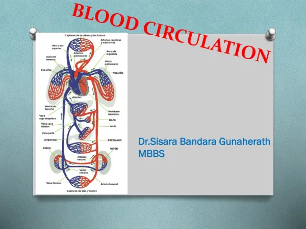
CEREBRAL BLOOD CIRCULATION DR JAMILA EL MEDANY
OBJECTIVES • At the end of the lecture, students should be able to: • List the cerebral arteries. • Describe the cerebral arterial supply regarding the origin, distribution and branches. • Describe the arterial Circle of Willis . • Describe the cerebral venous drainage and its termination. • Describe arterial & venous vascular disorders and their clinical manifestations.
CEREBRAL ARTERIAL SUPPLY A B • It is composed of two arterial systems: • A. Carotid System • B.Vertebro Basilar System
CAROTID SYSTEM It is composed of: The Anteior cerebral artery& Middle cerebral artery: both are branches of the Internal carotid artery
VERTEBRO BASILAR SYSTEM • The two Vertebral arteries ( from Subclavianartery) unite to form Basilar artery. • It divides at the upper border of the pons into two Posterior Cerebral arteries.
Distribution of the cerebral arteries on the superolateral surface of the cerebral H
Distribution of the cerebral arteries on the medial surface of the cerebral H
Anterior Cerebral Artery • It Supplies : orbital and medial surfaces of the frontal and parietal lobes • A narrow part on the superolateral surface.
Middle Cerebral Artery • It Supplies entire Superolateral surface: • Somatosensory Cortex • Motor Cortex • Language producing areas: • Broca's Area • Wernicke’s Area) • Auditory areas: • Primary auditory area • Auditory association (Heschl’sGyrus
Posterior Cerebral Artery • It Supplies: • Anterior and inferior parts of temporal lobe, Uncus, Inferior temporal gyri, • Inferior and Medial parts of Occipital lobe (visual areas)
CirculusArteriosus (of Willis) It joins the Carotid & Vertebrobasilar systems
It is locatedon the base of the brain • It encircles: • Optic Chiasma, Hypothalamus & pituitary gland Midbrain.
It is formed of: • 2Anterior cerebral arteries • 2Internal carotid arteries • 2 Posterior cerebral arteries • 2 Posterior communicating arteries • 1 Anterior communicating artery
Branches: • Perforating arteries (Anterior& Posterior): • Numerous small vessels that penetrate the surface of the brain through the anterior and posterior perforating substances. • APA supplies: • Large part of Basal Ganglia, • Optic chiasma, • Internal capsule & Hypothalamus • PPA supplies: • Ventral portion of Midbrain, parts of Subthalamus and Hypothalamus
Arterial Disorders • Stroke (Sudden occlusion • of the blood supply): • It can be: • Hemorrhagic • Ischemaic • Aneurysm • Angioma
EFFECT OF OCCLUSION of Cerebral arteries ACA MCA PCA
ACA • 1. Motor & sensory disturbances in the contralateral distal leg • 2. Difficulty in the Prefrontal lobe functions: • Cognitive thinking, Judgment, • Motor initiation and • Self monitoring
MCA • 1. Contralateral weakness of: • Face, Arm & Hand (more than leg) • 2.Contralateral sensory loss of: • Face, Arm & Hand (more than leg) 3. Visual field cut (damage to optic radiation) • 4. Aphasia (language disturbances ) • Broca's: production • Wernicke's: comprehension
PCA • 1. Visual disturbances • Contralateral homonymous hemianopsia • In Bilateral lesions: Cortical Blindness • patients unaware they cannot see (Anton's syndrome) • 2. Memory impairment • If the temporal lobe is affected
Cerebral Venous Drainage • Cortical Veins: • (A) Superficial • found in the Subarchnoid space Drain the cortical surfaces • (B) Deep veins: • Drain the deeper structures • These veins are thinwalled and devoid of valves. • They ultimately drain into the • Dural Venous Sinuses
Superficial Cortical Veins • 1. Superior cerebral veins (6 to 12) • Drain lateral surface of brain above the lateral sulcus • Terminate mainly into the Superior Sagittalsinus, and partly into Superficial middle cerebral vein. • 2. Inferior cerebral veins: • Run below the lateral sulcus • Drain the lateral surface of the temporal lobe • Terminate partly into superficial middle cerebral vein & partly into Transverse sinus.
3.Superficial middle cerebral vein: • Runs along the lateral sulcus • Terminates into the Cavernous sinus • It is connected posteriorlythrough Superior & Inferior anastomotic veins to Superior Sagittal& Transverse sinuses respectively.
Deep Cerebral Veins • Drain the internal structures (basal ganglia, internal capsule, thalamus) • They merge to form two Internal Cerebral Veins. • The two veins unite in the midline to form the Great Cerebral vein. • This short vessel is continuous with the Straight S
Dural Venous Sinuses Paired Single Superior sagittal. Inferior sagittal. Straight. Occipital. Transverse. Sigmoid. Cavernous. Petrosal Blood flows from transverse &sigmoid sinuses into IJV
Venous Disorders • Infarcation. • Sinus thrombosis: • (SSS thrombosis)can complicates ear infection . • Cavernous S thrombosis (as a complication of infection in the dangerous area of the face) • Obstruction of venous drainage of the brain leads to Cerebral swelling (edema)and raised ICP