Download
1 / 53
720 likes | 1.39k Views
TAKAYASU’S ARTERITIS. 1761-Morgagni 1830- Yamamoto 1905- Takayasu , prof of ophthal , presented the case of a 21 year old woman with characteristic fundal av anastamoses 1921-Shikhare-India 1951-Shimizu and Sano - summarized the c/f – pulseless disease. EPIDEMIOLOGY.

E N D
1761-Morgagni • 1830- Yamamoto • 1905- Takayasu, prof of ophthal, presented the case of a 21 year old woman with characteristic fundalavanastamoses • 1921-Shikhare-India • 1951-Shimizu and Sano - summarized the c/f –pulseless disease
EPIDEMIOLOGY • More case reports from Japan ,India, South-east Asia, Mexico • No geographic restriction • No race – immune • Incidence-2.6/million/year-N.America/Europe • The incidence in Asia is 1 case/1000-5000 women.
Age Mc-2nd & 3rd decade • May range from infancy to middle age • Indian studies-age 3- 50 yrs Gender diff • Japan-F:M=8-9:1 • India-F:M ratio varies from -1:1 - 3:1 ( Padmavati S, Aurora AP, KasliwalRR Aortoarteritisin India. J Assoc Physicians India 1987) • India=F:M- 6.4:1 (Panja et al, 1997 JACC)
Genetics • Japan - HLA-B52 and B39 • Mexican and Colombian patients - HLA-DRB1*1301 and HLA-DRB1*1602 • India- HLA- B 5, -B 21
Histopathology • Idiopathic c/c inflaarteritis of elastic arteries resulting in occlusive &/ ectatic changes • Large vessels, esp, Aorta & its main branches (brachiocephalic, carotid, SCL, vertebral, RA) • +Coronary & PA • Ao valve –usually not beyond IMA • Multiple segs with dis & skipped nl areas or diffuse involvement
Gross Histology Panarteritis-granulomatous lesion with giant cells a/c phase diffuse infil-mono granulomatousinfil 2)c/c phase-coll rich fibrous tissue- adventitia thicker than media 3)Healed phase-no infl cells, vas media scarred 1)Gelatinous plaques-early 2)White plaques-collagen 3)Diffuse intimal thickening Superficial– deep scarring circumferential stenosis 4)Mural thrombus 5)2⁰ atheromatous changes long standing, HTN
Wall thickening, Fibrosis, Stenosis, & Thrombus formation →end organ ischaemia • More a/c inflammation → destroys arterial media → Aneurysm (fibrosis inadequate) • Stenotic lesions predominate & tend to be B/L • Nearly all pts with aneurysms also have stenoses
Associated pathology-TB (LN)-55% Erthemamultiforme Bazinsdisease(erytinduratum) churgstrausssynd reteroperitoneal fib PAN,UC,CD etc
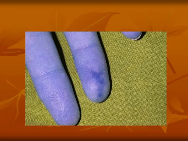
Clinical features Early pre pulseless/gen manif Late ischemic phase Sequel of occl of Ao arch/br Diminished/absent pulses (84–96%) Bruits (80–94%) Hypertension (33–83% ) RAS(28–75%) & CCF(28%) • Fever,weightloss,headache, fatigue,malaise,night sweats, arthralgia • +/_ splenomegaly/ cervical, axillarylymphadenopathy • Disappear partly/ completely in 3 months • 50% -no h/o acute phase
Coronary involvement in TA • Occurs in 10~30% • Often fatal • Classified into 3 types Type1:stenosis or occlu of coronary ostia Type2:diffuse or focal coronary arteritis Type3:coronary aneurysm
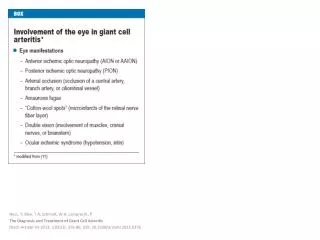
Occularinvolvement-Amaurosisfugax, pain behind eye, no real visual loss Hypertensive retinopathy Nonhypertensive retinopathy UYAMA & ASAYAMA CLASS stage 1- Dil of small vessels stage 2- Microaneurysm stage 3- Art-venanastomoses stage 4- Ocular complications Mild -stage 1 Moderate -stage 2 Severe -stages 3 & 4 Flourescienangio sensitive • Commonest • Arteriosclerotic –art narrowing, avnipping,silver wiring • Neuroretinopathy-exudates and papilloedema • Direct opthalmoscopy
PREGNANCY • Preg per se does not alter disease character • Compli of HTN mainly • Outcome usually favourable • Wong et al prog score for asses fetal outcome (inv AbAo+Ren art, MAP, time of onset –eclamp & treatment) • management of HTN essential • Meas of BP in UL –impossible/unreliable → oft more accurate in legs • HTN in 2nd stage Labour –risk for ICH-shortening stage . • pre-eclampsia, CCF, progressive RF,CVA • Fertility not affected
HTN is the most characteristic manifestation in Indian patients,suggesting a high frequency of lesions in the abdominal aorta, including the renal arteries, leading to renovascular hypertension
Maksimowicz-McKinnon K 2007 American College of Rheumatology
Ishikawa clinical classification of Takayasuarteritis 1978 4 Complications Retinopathy, Secondary HTN, AR, & Aneurysm
Cumulative survival • 5years -91% (event free survival -74.9%) • 10 years -84% (event free survival -64%) Single mild complication or no complication • 5 year event free survival 97% Single severe or multiple complications • 5 year event free survival 59.7% No deaths in groups I and IIA 19.6% mortality in groups IIB and III (CVA,CCF) Subramanyan R, Joy J, Balakrishnan KG, et al.SCT. Natural history of aortoarteritis (Takayasu’sarteritis). Circulation 1989; 80: 429-37.
Sharma BK, Jain S, Suri S, Numano F. Diagnostic criteria for Takayasuarteritis. Int J Cardiol 1996; 54 : S141-S147
Assessment of Disease Activity with Contrast-Enhanced MR Imaging YeonHyeonChoe et al j inter. radiology 2004 High-resolution contrast-enhanced T1-weighted spinechoMR small fields of view (14-20 cm) and thin slices(4-5 mm) • 26 patients with TA • Determinationof disease activity-concordantwith clinical findings (88.5%). • MR findings were concordant with lab findings -ESR 92.3% [24/26] andC-RP 84.6% [22/26]) MRI of Takayasu'sArteritis: Typical Appearances and Complications EijunSueyoshi et al August 12, 2005. J.int rad • provide almost all the anatomic informationneeded to enable early treatment • only technique needed for diagnosis of TA andits complications during the follow-up period.
Axial T1-weighted image- improvement of wall thickening of As Aoand PA after steroid therapy a/c phase-Axial T1-weighted image wall thickening of As aorta and PA
Findings of TA on MRI • mural thrombi • signal alterationswithin and surrounding inflamed vessels • vascular dilation • thickened aortic valvularcusps • multifocal stenoses • concentricthickening of the aortic wall • Disadvantages difficulty in visualizingsmall branch vessels and poor visualization of vascular calcification may falsely accentuate the degree of vascularstenoses (renal & subclavian)
[18F]fluorodeoxyglucose PET for diagnosingTakayasu’sarteritis • common [18F]FDG uptake pattern TA early phase -linear and continuous late phase-patchy rather than continuous ,linear • shown to identify more affected vascular regions than morphologic imaging with MRI • does not provide any information about changes in the wall structure or luminal blood flow • sensitivities of 83% and specificity 100% ( Meller Jet al. Value of F-18 FDG hybrid camera PET and MRI in earlyTakayasuaortitis.EurRadiol2003) • Sensitivity of 92%, specificity of 100% and a diagnostic accuracy of 94% ( Webb M et al. The role of 18F-FDG PET in characterising disease activity in Takayasuarteritis. EurJNucl Med Imaging 2004
Treatment of TA Control of vasculitis ・ Steroids If uncontrolled immunosuppressants: Cyclosporine,Cyclophosphamide, Mtx,Mycophenolatemofetil Symptomatic occlusion angioplasty/surgery thrombosis Anti-platelet therapy(low-dose Aspirin)
Medical treatment 0.7-1 mg/kg/day –prednisolone for 1-3 months common tapering regimen once remission ↓pred by 5 mg/week → 20 mg/day. Thereafter, ↓by 2.5 mg/week → 10 mg/day ↓1 mg/day each week, as long as disease does not become more active Pulse iv corticosteroids - CNS symptoms- no data to support
Steroids → 50% response • Methotrexate →further 50% respond • 25% with active disease will not respond to current treatments • resistant to steroids/ recurrent disease once corticosteroids are tapered cyclophosphamide (1-2 mg/kg/day), azathioprine (1-2mg/kg/day), or methotrexate (0.3 mg/kg/week) Mycophenolatemofetil/ anti TNF α agents- infliximab
Critical issue is in trying to determine whether or not disease is active • During Rx- regular clinical examination and ESR+ C-RP initially - every few days • CT or MR angio - 3 to 12 months - (active phase of Rx), and annually thereafter • Criteria for active disease
chronic phase- persistent inflammation steroidsshould be continued – <1.0mg/dL of s.C-RP and 20 mm/hof ESR
Surgical treatment • HTN with critical RAS • Extremity claudication limiting daily activities • Cerebrovascular ischaemia or critical stenoses of ≥3 cerebral vessels • Moderate AR • Cardiac ischaemia with confirmed coronary involvement • Aneurysms Recommended at quiescent state-avoids compli (restenosis, anastamotic failure, thrombosis, haemorrhage, & infection)
Surgical techniques • Carry high morbidity & mortality • Steno /aneurysm -anastomotic points • Progressive nature of TA • Diffuse nature of TA
Renal artery involvement • Best treated by PTA • Stent placement following PTA • Ostial lesions • Long segment lesions • Incomplete relief of stenoses • Dissection
ostial stenosis of the right renal artery after deployment of a stent
Renal PTA - 33 stenoses (20 pts) • Indi-sevHTN,angio 70% stenosis with pr grad 20mm, nl-ESR • Tech success -28 lesions (85%) clin success-14(82%) • Failures - Coexistent abdAo disease & tight, prox RAS • Tech diffi - tough, noncompliant stenoses, difficult to cross & resisted repeated, prolonged balloon inflations - backache & ↓SBP during balloon inflation • Follow-up –mean (8/12) -restenosis in 6 (21%) • Renal PTA in TA -tech difficulties; Short-term results - good, Complication rate-acceptable Sharma set al, AIIMS Am J Roentgenol. 1992 Feb;158(2):417-22
Aortoarteritic lesions Balloon dilation • safe & reasonably effective • Can be performed repeatedly without any added risks Balloon dilation diff from atherosclerotic lesions • Minimal intimal involvement –permits easy wiring and balloon crossing • Resistance to dilation – high fibrotic element in the stenotic lesion • restenosis> frequent in TA - diffuse and long stenotic lesions
Left subclavian angiograms- 95% stenosis with extensive collaterals Post angioplasty and stenting.
Joseph s et al, SCTJ VascIntervRadiol1994;5:573–580 Tyagi s et al, GB PantCardiovascInterventRadiol. 1998 May219-24 To compare PTA- Scl A in TA & athero 61 Scl A PTA (TA = 32 & athero = 23) PTA succ in 52 stenotis,3 occl TA -Higher balloon inflation P TA -more residual stenosis TA –restenosis more restnosis could be effectively redilated TA -Subclavian PTA - Safe, can be performed as effectively as in athero, good long-term results • PTA- Scl A in TA • 24 pts →26 Scl A VB insufficiency, UL claudication, or both • Aortography → (focal-14 ,< 3 cm,extensive-12) • Initial tech & clinical success – 81% (17 /19 steno,4/7occlu) • Follow-up → mean26 months → ISR -6 ( all ext) • Cumu patency –S/L-100/50% • Long-term results -excellent in focal lesions ,less durable extensive disease
Aortoplasty and Stenting • PTA -desc thoracic and/or abdAo (TA) stenosis • 16 pts (12+4)- HTN/severe b/l- LL claudication • Aortography – stenosis→ DTA-5, abd Ao-10, Both -1 • Initial tech & clinical success -100% • patency rate of 67%in a 52-month follow-up • Follow-up (mean 21months)- Restenosis -3 • PTA has a definite role in TA management • residual gradient < 20 mm -criterion for successful aortoplasty • long-segmentdisease, dissection or persistence of a grad > 20 mm Hg after PTBA- aortic stenting Rao AS et al, SCT Radiology. 1993 Oct;189(1):173-9
long-segment diffuse stenotic involvement of the DTA after deployment of stents.
Treatment for cor A occulusion in TA Surgery (CABG)- often not indicated ・IMA can’t be used often • occlu of Innomi A / Scl A • calcification of aorta High incidence of restenosis:36% Angioplasty(PTCA) ・alternative to surgery Very high incidence of restenosis:78% DES-effectiveness ?
Percutaneous Management of Aneurysmal Lesions • Aneurysmaldilatation- isolation or together with stenoticlesions • fusiform or saccular • one of the major complicationsrelated to the prognosis in TA • Incidence of aneurysm rupture -low • Management -mainly surgical. • Covered stent-grafts may be useful