Download
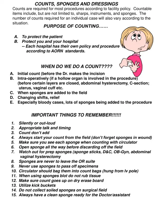
1 / 36
370 likes | 577 Views
Casts, Drains and Dressings. Fracture management stabilization devices. External fixation Casts Plaster (fast, medium, slow-setting) Fiberglass Types Shoulder spica Minerva jacket Body cast Short arm/leg Long arm/leg Hip spica Cylinder cast. Minerva Jacket.

E N D
Fracture management stabilization devices • External fixation • Casts • Plaster (fast, medium, slow-setting) • Fiberglass • Types • Shoulder spica • Minerva jacket • Body cast • Short arm/leg • Long arm/leg • Hip spica • Cylinder cast
Minerva Jacket • A plaster of Paris body cast incorporating the head and trunk, usually for fracture of the cervical spine.
Body cast • Often not a true one piece cast. • What people call a full body cast is often a combination of many smaller casts around a Hip Spica or a Minerva Jacket.
Goals of Casting/Splinting • Relieve pain • Augment healing • Stabilize fracture • Prevent further injury • Splinting better if practical because it is easier to manage swelling considering the entire limb is not isolated by a circumferential cast
Casting Considerations • Casts • Proper placement of cast brings patient safety issues • Patient’s limb should be elevated • Webril should be placed so no wrinkles are in cotton to cause pressure sores • As plaster or fiberglass is placed, assistant must not make marks in plaster as it dries—these may cause pressure sores • Reflective materials will reflect heat given off by casting material if fiberglass and may burn patient’s limb • Tip of limb should be cleaned of all prepping solution so patient may be monitored for signs of circulatory disruption: increasing pain, pain that progresses into numbness, cyanotic skin, cold skin, poor capillary refill
Drains • Drains are inserted to: • Evacuate collections of pus, blood or other fluids • Drain potential collections
Drains • Arguments for their use include: • Drainage of fluid removes potential sources of infection • Drains guard against further fluid collections • May allow the early detection of anastomotic leaks or hemorrhage • Leave a tract for potential collections to drain following removal
Drains • Arguments against their use include: • Presence of a drain increases the risk of infection • Damage may be caused by mechanical pressure or suction • Drains may induce an anastomotic leak • Most drains abdominal drains infective within 24 hours
Drains • Drains can be: • Open or closed • Active or passive – more important • Drains are often made from inert silastic material • They induce minimal tissue reaction
Open drains • Open drains • Include corrugated rubber or plastic sheets • Drain fluid collects in gauze pad or stoma bag • They increase the risk of infection
Closed drains • Consist of tubes draining into a bag or bottle • They include chest and abdominal drains • The risk of infection is reduced
Active drains • Active drains are maintained under suction • They can be under low or high pressure
Passive drains • Passive drains have no suction • Function by the differential pressure between body cavities and the exterior
Examples of drains and operations • Plastic surgery including myocutaneous flap surgery • Breast surgery (to prevent collection of blood and lymph) • Orthopedic procedures (associated with greater blood loss) • Chest drainage • Chest surgery (with for example the associated risks of raised intrathoracic pressure and tamponade)
Examples of drains and operations • Pancreatic surgery (to drain secretions) • Biliary surgery • Thyroid surgery (concern over hematoma and hemorrhage around the airway) • Neurosurgery (where there is a risk of raised intracranial pressure) • Nasogastric tubes • Urinary catheters
Nasogastric tubes • Following abdominal surgery gastointestinal motility is reduced for a variable period of time • Gastrointestinal secretions accumulate in stoma and proximal small bowel • May result in: • Postoperative distension and vomiting • Aspiration pneumonia • Little clinical evidence is available to support the routine use of nasogastric tubes • May increase the risk of pulmonary complications • Of proven value for gastrointestinal decompression in intestinal obstruction • Tubes are usually left on free drainage • Can be also aspirated maybe every 4 hours • Can be removed when volume of nasogastric aspirate is reduced
Urinary catheters • A urinary catheter is a form of drain • Commonly used to: • Alleviate or prevent urinary retention • Monitor urine output • Can be inserted transurethrally or suprapubically
Urinary catheters • Catheters vary by: • The material from which they are made (latex, plastic, silastic, teflon-coated) • The length of the catheter (38 cm 'male' or '22 cm 'female') • The diameter of the catheter (10 Fr to 24 Fr) • The number of channels (one, two or three-irrigation) • The size of the balloon ( 5ml to 30 ml) • The shape of the tip – i.e. Coude tip
Do's and don'ts of urinary catheters • Choose an appropriate sized catheter • Insert using an aseptic technique • Never insert using force • Do not inflate the balloon until urine has been seen coming from the catheter • Record the residual volume • Do not use a catheter introducer unless you have been trained in its use • If difficulty is encountered inserting a urinary catheter consider a suprapubic • Remove at the earliest possibility
Types of Active Drains • JP’s • Hemovac’s • Pleuravac
JP’s • Can be flat or round • Will always have a “grenade” – the bulb section. • The bulb provides the suction, thus making it an active drain.
Hemovac’s • Used post-op; • for up to 400mL of drainage; • uses negative pressure by spring action. • Springs are inside the drain container.
Pleur-vac • Chest drainage units are used after open heart, thoracic or emergency surgery procedures to evacuate air and fluid present in the thoracic cavity. Without chest drainage, positive pressure may build up in the thoracic cavity and lead to significant patient problems. • Pleur-evac inventor and surgeon, Dr. Sidney Mishkin, was inspired to act after he saw a nurse drop and break glass bottles traditionally used in chest drainage. • Look to BB for further information – Chest Tubes.docx
Pleur-vac • This is the new system! • Much improved. • Chest tubes hook up to the large tubes in the top of the active drain chamber. • Another tube hooks up to regulated wall suction
Types of PassiveDrains • Penrose • Cigarette • T-Tube • Gastrostomy Tube • Cystostomy tube • Nephrostomy tube
Penrose • A Penrose drain is a surgical device placed in a wound to drain fluid. • It consists of a soft rubber tube placed in a wound area, to prevent the build up of fluid.
Cigarette Drain • A drain made of gauze surrounded by a rubber tissue, rubber dam, or rubber tubing. • Encourages draining by wicking action
T-Tube • Placed within the biliary system. • The T-tube allows bile to drain out of the patient's body into a small pouch, known as a bile bag.
Gastrostomy Tube • used to provide nutrition to patients who cannot obtain nutrition by swallowing. • Also used to remove gastric contents. • Also called a feeding tube • a tube inserted through a small incision in the abdomen into the stomach
Cystostomy tube • the construction of an artificial opening from the bladder through the abdominal wall, permitting the drainage of urine.
Nephrostomy tube • Tube inserted into the kidney to drain urine. • The ureter is the fibromuscular tube that carries urine from the kidney to the bladder. When this tube is blocked, urine backs up into the kidney. • Serious, irreversible kidney damage can occur because of this backflow of urine. Infection is also a common consequence in this stagnant urine.
Drain considerations • General guidance • If it is an active drain, the drain can be attached to a suction source (and set at prescribed pressure). • Ensure the drain is secured (dislodgement is likely to occur when transferring patients after anesthesia). Dislodgement can increase the risk of infection and irritation to the surrounding skin. • Accurately measure and record drainage output. • Monitor changes in character or volume of fluid. Identify any complications resulting in leaking fluid (particularly for example bile or pancreatic secretions) or blood. • Use measurements of fluid loss to assist intravenous replacement of fluids.
Dressings • Look at ST 270-272