Pulmonary Embolism
Pulmonary Embolism. Dr. Meg- angela Christi Amores. Venous Thromboembolism (VTE). Deep Vein Thrombosis (DVT) Pulmonary Embolism (PE). Pulmonary Embolism (PE). Pathophysiology Embolization Venous thrombi dislodge Enters the pulmonary circulation


Pulmonary Embolism
E N D
Presentation Transcript
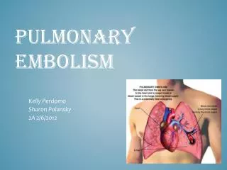
Pulmonary Embolism Dr. Meg-angela Christi Amores
Venous Thromboembolism (VTE) • Deep Vein Thrombosis (DVT) • Pulmonary Embolism (PE)
Pulmonary Embolism (PE) • Pathophysiology • Embolization • Venous thrombi dislodge • Enters the pulmonary circulation • Or paradoxically, to arterial circulation
Pathophysiology • Physiology • most common gas exchange abnormalities are hypoxemia (decreased arterial PO2) • inefficiency of O2 transfer across the lungs • Increased pulmonary vascular resistance • Impaired gas exchange • Alveolar hyperventilation • Increased airway resistance • Decreased pulmonary compliance
Pathophysiology • Right Ventricular Dysfunction • Progressive right heart failure is the usual cause of death from PE • RV contraction continues even after the left ventricle (LV) starts relaxing • the interventricular septum bulges into and compresses an intrinsically normal left ventricle
Diagnosis • Clinical Evaluation • Nonspecific signs and symptoms • Known as “the Great Masquerader” • most frequent history is unexplained breathlessness • Dyspnea • Tachypnea • dyspnea, syncope, hypotension, or cyanosis • pleuritic pain, cough, or hemoptysis
Diagnosis • Laboratory • Blood tests: D dimer assay • Elevated cardiac markers: Troponin • ECG: S1Q3T3 sign: • an S wave in lead I, Q wave in lead III, and inverted T wave in lead III • T-wave inversion in leads V1 to V4
Diagnosis • Imaging • Venous Ultrasound • Chest XRay: • Westermark's sign - focal oligemia • Hampton's hump - a peripheral wedged-shaped density above the diaphragm • Palla’s sign - an enlarged right descending pulmonary artery • Chest CT Scan with contrast • Lung Scan
Treatment • Anticoagulation • foundation for successful treatment • parenteral drug: unfractionated heparin (UFH), low molecular weight heparin (LMWH), or fondaparinux • "bridge" to stable, long-term anticoagulation with a vitamin K antagonist : WARFARIN
Treatment • IVC filter • Maintain adequate circulation • Fibrinolysis • Pulmonary Embolectomy • Pulmonary Thromboendarterectomy • Emotional Support