Download
1 / 15
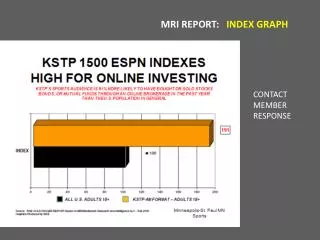
180 likes | 613 Views
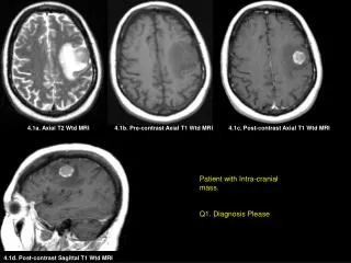
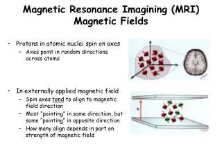
Magnetic Resonance Imagining (MRI) Magnetic Fields. Protons in atomic nuclei spin on axes Axes point in random directions across atoms In externally applied magnetic field Spin axes tend to align to magnetic field direction

E N D
Magnetic Resonance Imagining (MRI) Magnetic Fields • Protons in atomic nuclei spin on axes • Axes point in random directions across atoms • In externally applied magnetic field • Spin axes tend to align to magnetic field direction • Most “pointing” in same direction, but some “pointing” in opposite direction • How many align depends in part on strength of magnetic field
MRI Magnetic Fields Cont’d • In an externally applied magnetic field, atomic nuclei with an odd number of nucleons (protons + neutrons) precess • = Spin axis wobbles • Rate determined both by properties of the atom • And by the strength & homogeneity of the magnetic field • Hydrogen atoms in a 1.5 Tesla field precess at ~64 MHz = Radio frequency [rf] • Timing (phase) of wobble still random across atoms
Resonance • A radio frequency pulse (rf pulse) of a particular frequency is applied to the aligned protons in the magnetic field • When rf pulse frequency = precession frequency = Larmor frequency, nuclei resonate to it • Pushes spinning protons into phase with one another • Amplitude of wobble of the whole magnetic field generated by the spinning protons increases (= spin axis is pushed farther out) • Increases “transverse component” of magnetic field • Which is what’s measured • How far spin axis moves (= flip angle) depends on rf pulse intensity and duration
Why is this useful? • Takes characteristic amount of time after rf pulse for protons: • To get back out of phase (= de-phase) • And to settle back to original wobble amplitude in fixed field • Depending on what other kinds of atoms are nearby • i.e, the kinds of molecules the atoms are in, in part • As protons settle back into alignment with fixed field, the strength of the magnetic field they generated decreases • Variations in how long it takes the protons to de-phase & to settle back to original wobble amplitude in fixed field • Can be used to distinguish among different substances
How does this make IMAGING possible? • How know where magnetic field being measured comes from?) • Use gradient magnetic fields (Lauterbur Nobel Prize) • Generate field with gradation in field strength with only a small region at 1.5 Tesla • Only hydrogen atoms within that region respond to 64 MHz pulse • So, response must come from protons within 1.5 T region • Keep moving position of 1.5 T area to localize source of responses to repeated rf pulses • Size of 1.5 T region determines granularity of localization of response
Fixed field magnet is (almost) always on! • - Child killed in 2001 at Westchester • Medical Center when an oxygen tank • brought into magnet room was pulled • into center of magnetic field • In 2000, police officer’s gun pulled • from his hand into magnet & discharged • a bullet into the wall on the way in
Structural MRI • Anatomical scans generally measure hydrogen atoms in water • Since different kinds of tissue have different proportions of water • Typical anatomical scan voxel granularity = 1 x 1 x 3-5 mm
Functional MRI (fMRI) • Substance measured is hemoglobin (iron) in blood • Blood flow increases to active brain regions • Increases more than is usually needed • So ratio of de-oxygenated to oxygenated blood decreases • Oxygenated & de-oxygenated hemoglobin respond differently to magnetic field and rf pulses • Use this to detect where more oxygenated blood goes after some event • Takes several seconds for the response to peak • Timing seems to vary some across different brain regions • Fastest reliably detectable pre-peak response so far = 2 - 4 sec • Signal strength change very small – generally less than 1% change • If signal strength large, probably due to draining vein
Important to avoid measuring this instead of this
fMRI Cont’d • Spatial resolution: • Ultimate limit probably spatial specificity of the circulatory system • Worse than structural MRI • Typical functional scan voxels = 3 x 3 x 5 mm • Temporal resolution: • IF blood flow is what’s measured, never going to be faster than seconds • Working on detecting the brief initial decrease in oxygenated blood preceding increased blood flow • Working on imaging other substances
fMRI Data Analysis • Much of the brain is active much of the time • (e.g., “default network”) • Try to isolate regions that are specific to some aspect of the event of interest • One widely used approach is to look for voxels whose time course of signal strength change after stimulus is correlated with an idealized hemodynamic response function • Decide on a threshold for correlation strength and only further analyze voxels exceeding that threshold
Subractive Logic • Construct 2 conditions that you believe differ in just 1 important way • Treat one as baseline & subtract it from the other to get rid of all the • activity the 2 have in common • And then analyze what’s left • May only analyze part of what’s left because may threshold (again), • and/or may only analyze Regions of Interest (ROI) • These are all ways to cut down the number of statistical comparisons done
Subtractive Logic, Cont’d • Similar logic used in comparing conditions in most other kinds of experiments, too • But there’s been a very unfortunate tendency in much of the imaging literature so far, • For researchers who don’t have a good understanding of the many ways that different kinds of stimuli and/or tasks and/or situations can differ, • To claim that they’ve located “phonological word processing”, or “irregular morphological inflection processing”, or some such aspect of language processing • When other confounded differences between conditions are equally good candidates (such as plain old difficulty) for explaining the effects