Download

1 / 18
180 likes | 234 Views
Learn about the physiology of veins and how skeletal muscles support blood flow, leading to conditions like varicose veins. Explore risk factors, morphology, complications, causes, and clinical syndromes associated with venous thrombosis. Discover lymphangitis and lymphedema, including primary and secondary causes.

E N D
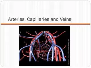
Normal vein physiology Skeletal muscles Skeletal muscles They “pump” the blood towards the heart and prevent backflow of blood by compressing the veins when contracted. Prevent the backflow of blood
Because of the difference in tunica media between the arteries and veins, the arteries are stronger and the veins are easier to compress and dilate (the vein is weaker and tortuous)
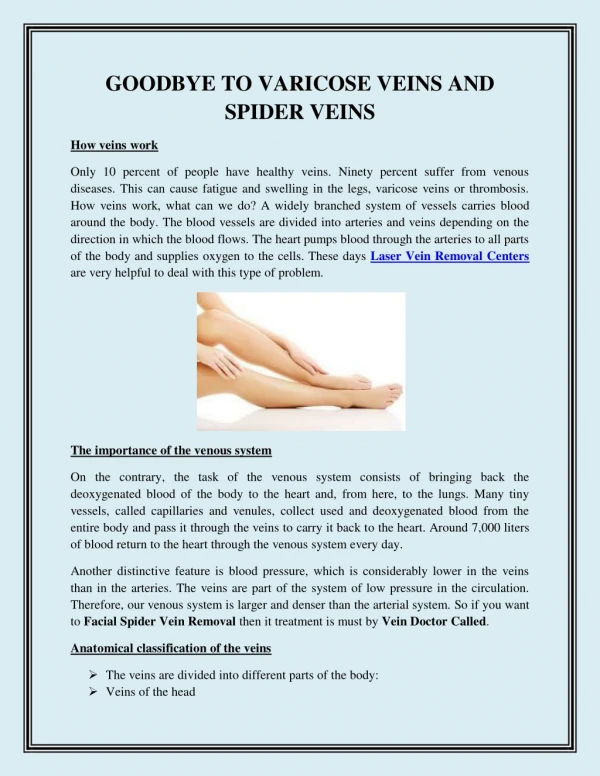
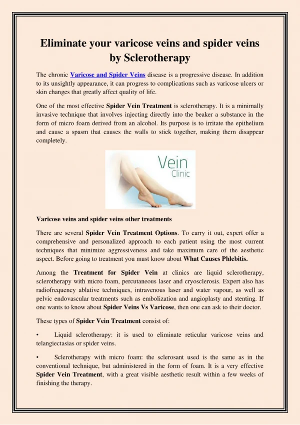
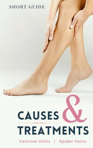
Veins and lymphatics • Varicose Veins • are abnormally dilated, tortuous veins produced by prolonged increase in intra-luminal pressure, loss of vessel wall support (weakness of veins) or abnormality in pocket valves. • The superficial veins of the leg are typically involved
Varicose Veins They appear because of gravity After standing up for 2 hours At resting state
venous pressures in legs can be markedly elevated venous stasis and pedal edema (simple orthostatic edema). • Some 10% to 20% of adult males and 25% to 33% of adult females develop lower extremity varicose veins females have higher susceptibility Because of 1-pregnancy 2-weaker muscles less “pumping” 3- estrogens by decreasing the overall muscle tone.
Risk factors • Obesity increases intraluminal pressure • Female gender less muscles and high estrogen • Pregnancy compressing the pelvic veins. • Familial tendency (premature varicosities results from imperfect venous wall development making the walls weaker)
Morphology (microscopic) • wall thinning • intimal fibrosis in adjacent segments • spotty medial calcifications (phlebosclerosis) • Focal intraluminal thrombosis • venous valve deformities (rolling and shortening)
complications The problem is usually only cosmetic • stasis, congestion, edema, pain, and thrombosis • chronic varicose ulcers in the skin • embolism is very rare. causes
Thrombophlebitis And Phlebothrombosis Sometimes the inflammation occurs first and sometimes thrombosis • interchangeable terms • = (Inflammation + thrombosis) of veins • The deep leg veins account for more than 90% of cases • the most important clinical predispositions are: congestive heart failure, neoplasia, pregnancy, obesity, the postoperative state, and prolonged bed rest or immobilization All causes stasis
Thrombophlebitis of upper limb veinsare usually associated with local risk factors like: catheter or canulasite (where inflammation sometimes occur); or in some cases can be associated with systemic hypercoagulabilities. • local manifestations: distal edema, cyanosis, superficial vein dilation, heat, tenderness, redness, swelling, and pain • Distant manifestations: emboli
Migrates between veins • Clinical syndromes associated with venous thrmobosis: 1- Migratory thrombophlebitis (Trousseau sign): hypercoagulability occurs as a paraneoplastic syndrome related to tumor elaboration of pro-coagulant factors - Most often related to GI carcinomas ( colon . Stomach and pancreatic cancers)
2- The superior vena caval syndrome • caused by neoplasms (most commonly lung cancer)that compressorinvade the superior vena cava. • A characteristic clinical complex including marked dilation of the veins of the head, neck, and arms (upper limb)with cyanosis , edema , congestion… (all the local manifestations).
3- The inferior vena caval syndrome • can be caused by neoplasms that compress or invade the inferior vena cava (IVC)- particularly hepatocellularcarcinoma and renalcellcarcinoma, which show a striking tendency to grow within veins- • induces marked lower extremity/limb edema, distention of the superficial collateral veins of the lower abdomen, and-with renal vein involvement-massive proteinuria.
Lymphangitis • is the acute inflammation due to bacterial infections spread into the lymphatics • most common are group A β-hemolytic streptococci. • lymphatics are dilated and filled with an exudate of neutrophils and monocytes. • Presentation :red, painful subcutaneous streaks(the inflamed lymphatics),with painful enlargement of the draining lymph nodes (acute lymphadenitis) (when the infection reachsthe lymph nodes). • Its usually localized but Sometimes subsequent passage into the venous circulation can result in bacteremia or sepsis(specially in immunocompetent patients).
lymphedema Edema caused by a problem in lymphatics • can occur as: 1- Primary (A congenital defect early in life),resulting from lymphatic agenesis(failure to form)or hypoplasia (less than normal). 2- Secondary or obstructive lymphedemamore common • 1- blockage of a previously normal lymphatic; e.g. Malignant tumors • 2- Surgical procedures that remove • lymph nodes ex. Breast cancer and the • edema will occur in the upper limb • 3- Post-irradiation (e.g. breast cancer) • 4- Fibrosis • 5- Filariasisinfection by filarial parasite • that leads to blockage of lympatics • 6- Post-inflammatory thrombosis and scarring
chylous Accumulation of lymph outside the lymphatics (Means milky substance) • Milky accumulations of lymph in various body cavities • caused by rupture of dilated lymphatics, typically obstructed secondary to an infiltrating tumor mass (meaning a connection between a lymphatic and a body cavity occured) - chylousascites(abdomen) • Chylothorax (chest) • Chylopericardium (pericardium) Special thanks to