Download
1 / 19
410 likes | 2.43k Views
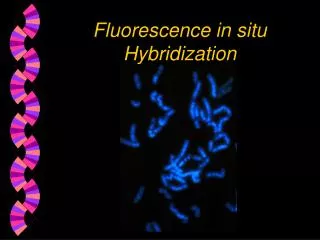
Fluorescence in situ Hybridization. Fluorescence in situ Hybridization (FISH). FISH - a process which vividly paints chromosomes or portions of chromosomes with fluorescent molecules Opening picture - Human M-phase spread using DAPI stain. Fluorescence in situ Hybridization (FISH).

E N D
Fluorescence in situ Hybridization (FISH) • FISH - a process which vividly paints chromosomes or portions of chromosomes with fluorescent molecules • Opening picture - Human M-phase spread using DAPI stain
Fluorescence in situ Hybridization (FISH) • Identifies chromosomal abnormalities • Aids in gene mapping, toxicological studies, analysis of chromosome structural aberrations, and ploidy determination
Fluorescence in situ Hybridization (FISH) • Used to identify the presence and location of a region of DNA or RNA within morphologically preserved chromosome preparations, fixed cells or tissue sections
Fluorescence in situ Hybridization (FISH) • This means you can view a segment or entire chromosome with your own eyes • Was often used during M phase but is now used on I phase chromosomes as well
Fluorescence in situ Hybridization (FISH) • Advantage: less labor-intensive method for confirming the presence of a DNA segment within an entire genome than other conventional methods like Southern blotting
FISH Procedure • Denature the chromosomes • Denature the probe • Hybridization • Fluorescence staining • Examine slides or store in the dark
FISH Uses • Detection of high concentrations of base pairs • Eg: Mouse metaphase preparation stained with DAPI (a non-specific DNA binding dye with high affinity for A-T bonds)
FISH Uses • Centromere regions stained brighter - means they are rich in A-T bonds • Also used in germ cell or prenatal diagnosis of conditions such as aneuploidies
FISH and Telomeres • Telomeric probes define the terminal boundaries of chromosomes (5’ and 3’ ends) • Used in research of chromosomal rearrangements and deletions related to cell aging or other genetic abnormalities
FISH and Telomeres • Special telomeric probes specific to individual chromosomes have been designed • Probe is based on the TTAGGG repeat present on all human telomeres
FISH and Telomeres • Application in cytogenetics - can detect submicroscopic deletions and cryptic translocations of genes associated withunexplained mental retardation and miscarriages
FISH and Telomeres - Medical from TelVysion DNA website • “A translocation between chromosomes 12 and 21 with breakpoints at bands 12p13 (TEL)[telomere] and 21q22 (AML1) occurs in at least 25 percent of childhood B-cell acute lymphocytic leukemia”
FISH and Telomeres - Medical • The “LS1 TEL/AML1 ES Dual-Color Translocation Probe” is designed to detect the TEL and AML1 gene fusion • This fusion is undetectable by standard cytogenetics, but can be seen with FISH
FISH - Medical • FISH can be used in the study of transgenic animals (eg: Polly) • Selective markers show if the human DNA was inserted successfully and pinpoint where the human DNA is • Transgenic Mouse