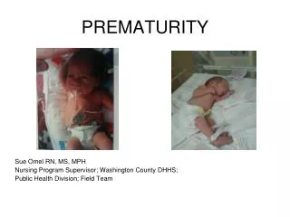

Prematurity
E N D
Presentation Transcript
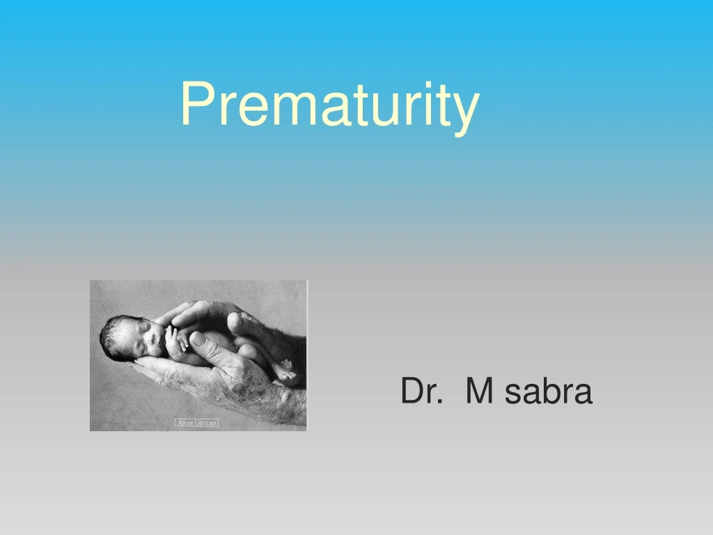
Prematurity Dr. M sabra
Definitions Post-conceptional age: gestational age plus post-natal age premature: gestational age less than 37 weeks or 259 days moderately premature: 31-36 weeks severely premature: 24-30 weeks 5-10% of live births High morbidity and mortality due to immature organ systems Responsible for 75% of perinatal deaths
postterm: greater than 41 weeks low birth weight (LBW): < 2,500 Gm (only a bit over half of LBW infants are premature)very low birth weight (VLBW): < 1,500 Gm newborn: first day of life neonate: first month of life infant: first year of life
Immediate/early complications hypoxia/ischemia intraventricular hemorrhage sensorineural injury respiratory failure necrotizing enterocolitis cholestatic liver disease nutrient deficiency social stress
Early complications and associated sequelae Hypoxia/ischemia mental retardation spastic diplegia microcephaly seizures poor school preformance
Early complications and associated sequelae Nutrient dificiency osteopenia fractures anemia vitamin E deficiency growth failure
Early complications and associated sequelae Social stress child abuse/neglect failure to thrive
Special considerations Respiratory breathing may initially be exclusively nasal spontaneous neck flexion may cause airway obstruction and apnea diaphragm is most important respiratory muscle maternal betamethasone or dexamethasone 48 hours before delivery increases surfactant production and decreases mortality after 30 weeks gestation
Organ system complications Respiratory respiratory distress syndrome (RDS) bronchopulmonary dysplasia pneumothorax pneumomediastinum congenital pneumonia pulmonary hypoplasia pulmonary hemorrhage apnea
Respiratory distress syndrome Respiratory distress syndrome (RDS) results from immaturity of the lungs, particularly the production of surfactant in the premature infant. RDS is typified by tachypnoea, dyspnoea, cyanosis and grunting’, non-compliant lungs, widespread atelectasis on chest radiograph and the presence of hyaline membranes in terminal airways (previous terminology: hyaline membrane disease). may be complicated by air leak (pneumothorax, pneumomediastinum, pulmonary interstitial emphysema) and lead to bronchopulmonary dysplasia (BPD) and chronic lung disease .
Nasal continuous positive airway pressure (nCPAP) is the method of choice for ventilatory support ( increase in FRC, reduce atelectasis and improve work of breathing). Babies are extubated to nCPAP as soon as possible. Intermittent positive pressure ventilation (IPPV) increases the risk of BPD and is used only if necessary (e.g. for the administration of surfactant, for VLBW infants at <28 weeks, and if oxygenation with nCPAP is inadequate with a fraction of inspired oxygen (FiO2) >50e60%). The use of strategies to allow permissive hypercapnia may also reduce the risk of ventilator-induced injury Different ventilation modes are used to reduce volu/barotrauma. These include various forms of patient-triggered ventilation (such as pressure support and proportional assist ventilation) and minimization of excessive tidal volumes (volume targeted ventilation and high frequency oscillation). . Steroids are no longer used to facilitate weaning from long-term ventilation.
bronchopulmonary dysplasia BPD is defined as oxygen dependency for more than 28 days after birth and an abnormal chest radiograph. The chest radiograph changes in early BPD may be indistinguishable from the ground glass appearance of RDS. But later radiographs show patchy atelectasis and cystic changes with hyperexpansion and areas of emphysema.
Apnoea of prematurity Apnoea is a pause in breathing of more than 20 seconds or one of less than 20 seconds associated with bradycardia and/or cyanosis. Apnoea of prematurity is commonly seen in neonatal units, Classified : central (brainstem, peripheral chemoreceptor immaturity), obstructive (reduced tone, asynchrony of diaphragm/upper airway activity, excessive neck flexion, structural abnormalities) or mixed cause.
In adult life, hypoxia and hypercapnia increase ventilation. Premature and newborn term babies respond to hypoxia by a brief increase in ventilation followed by apnea and have a blunted response to hypercapnia. In the term infant, normal responses to hypercapnia and hypoxia are seen by 3 weeks of age, but this is delayed in premature infants. Apnea in premature infants is exacerbated by hypoxia, sepsis, intracranial hemorrhage, metabolic abnormalities, hypo/hyperthermia, upper airway obstruction, heart failure, anemia, vasovagal reflexes and drugs, including prostaglandins and anaesthetic agents. Apnoeas are treated by stimulation, bag-mask ventilation, addressing underlying abnormalities, the use of respiratory stimulants such as caffeine or aminophylline, nCPAP or ventilation. Term neonates are at low risk of postoperative apnoea after routine minor surgery at 44 weeks post-conception. However, in premature neonates the probability of postoperative apnoeas decreases to less than 1% only at 60 weeks post-conception
Organ system complications Cardiovascular Cardiac output relatively dependent on heart rate Immature sympathetic innervation patent ductus arteriosus (PDA) hypotension hypertension bradycardia (with apnea) congenital malformations
Patent ductus arteriosus The arterial duct is one of the fetal shunts and is closed in 3e4 days in 90% of term and ‘well’ premature babies in response to a rise in oxygen tension after birth and a fall in circulating prostaglandins. PDA is seen in 50% of VLBW infants due to low oxygen tension,continuing high prostaglandin levels, or abnormal stimuli such as acidosis and expansion of the circulating volume. Aorto-pulmonary shunting though the PDA causes high pulmonary blood flow, worsening RDS, cardiac failure and low diastolic pressure. PDA is a risk factor for intraventricular haemorrhage, necrotizing enterocolitis and CLD. .
PDA typically becomes symptomatic at 5e10 days as pulmonary vascular resistance falls; it presents with worsening respiratory function, bounding pulses, a continuous murmur and chest radiograph that shows cardiomegaly and increased lung shadowing. Diagnosis is confirmed by echocardiography. Conventional treatment for symptomatic PDA is fluid restriction, diuretics (furosemide) or medical closure with indomethacin or ibuprofen. Non-steroidal anti-inflammatory drugs (NSAIDs) may worsen renal function, and have been associated with gastrointestinal haemorrhage and perforation These agents are contraindicated in the presence of thrombocytopenia. Surgical closure of symptomatic PDA is indicated for failed medical treatment or when NSAIDs are contraindicated
Special considerations Nervous system • Brain has 2 growth spurts • neuronal cell multiplication 15-20 weeks gestation • 2. glial cell multiplication 25 weeks to 2 years of life • Blood vessels more fragile increased risk of intracerebral hemorrhage • Developmental aspects of pain perception • Pain pathways develop during the second and third trimester. By 26 weeks’ gestation premature neonates respond to tissue damage by withdrawal reflexes and activation of the stress response. Pathways between the thalamus and somatosensory cortex function by 29 weeks’ gestation. • The precise gestational age when a neonate is able to perceive pain is unknown.
Organ system complications Neurologic intraventricular hemorrhage periventricular leukomalacia hypoxic-ischemic encephalopathy seizures retinopathy of prematurity (retrolental fibroplasia) hypotonia congenital malformations kernicterus (bilirubin encephalopathy)
Intraventricular haemorrhage 21% of babies born at less than 25 weeks’ gestation have severe disability A major determinant of cerebral impairment is IVH, particularly complicated by ventricular enlargement, parenchymal infarction or cystic periventricular white-matter injury. Major IVH usually occurs within the first few days of life and is detected by cranial ultrasound. Factors shown to increase the incidence of IVH or later neurodevelopmental delay include RDS, hypotension or fluctuating blood pressure, the use of hypertonic infusions and aggressive volume expansion. The normal lower limit MAP is roughly equivalent to the gestational age on the first day of life, with a MAP of at least 30 mmHg for all infants by day 3 of life. Management of hypotension requires judicious use of volume expansion (crystalloid or colloid) and the early use of inotropic agents such as adrenaline, dopamine or dobutamine. Aggressive volume expansion should be avoided, especially in the first few days of life.
Periventricular leukomalacia 12-25% of LBW infants with increase risk of mental handicap describes histological changes in periventricular white matter seen in premature infants. The pathogenesis of periventricular leukomalacia is associated with hypoxic-ischaemic or toxic injury, infection, impaired cerebral autoregulation, cerebral ‘steal’ due to a large PDA and severe hypocarbia. Bilateral occipital cystic periventricular leukomalacia is a very strong predictor of cerebral palsy
Retinopathy of prematurity and oxygen toxicity Retinopathy of prematurity (ROP) is seen in LBW infants less than 32 weeks’ gestation. Hyperoxia in the first weeks of life causes vasoconstriction of retinal vessels, which leads to retinal ischaemia and a subsequent vasoproliferative phase. Good neonatal care, ophthalmic screening and treatment can prevent ROP. There is concern that even brief exposure to high oxygen levels is associated with increased morbidity and mortality in VLBW infants; fluctuations in oxygen levels should be avoided nd oxygen saturation maintained between 88e95%, not exceeding 95%. Newborn resuscitation should be carried out with room air rather than 100% oxygen.
Organ system complications Hematologic anemia hyperbilirubinemia (direct or indirect) subcutaneous or organ hemorrhage (liver, adrenal) disseminated intravascular coagulopathy vitamin K deficiency hydrops
Organ system complications Gastrointestinal necrotizing enterocolitis poor gastrointestinal function or motility
Necrotizing enterocolitis NEC occurs mainly in preterm infants, with an incidence of about 7% and a mortality of 15-30%. It has a multifactorial aetiology ( prematurity and poor mucosal integrity, hypoxia, early feeding with formula milk and colonization with pathogenic bacteria) NEC causes inflammation and transmural necrosis and can affect any part of the intestine, typically the terminal ileum, caecum and ascending colon. The classical presentation is of abdominal distension, bloody stool and bile-stained aspirates, but signs of sepsis may predominate, with vague non-specific signs progressing to apnoea with shock and DIC. Intestinal perforation may cause a localized mass and the abdominal wall may be reddened in the presence of peritonitis. NEC is associated with pneumatosis (gas within the bowel wall), and a characteristic appearance on a radiograph of dilated thickened loops of bowel with intramural gas. Free gas may be visible on horizontal shoot-through in the presence of a perforation.
Investigations may also reveal low platelet count, raised C-reactive protein and metabolic acidosis. Babies with severe disease may have exposed T antigen on red blood cells, which leads to haemolysis in the presence of transfused blood products containing anti-T antibodies. Blood products with low-titre anti-T antibodies will be required for T-antigen-positive infants (packed cells reconstituted in SAG-M are safe). ( saline-adenine-glucose-mannitol ; 376 mOsm/l) Treatment of NEC consists of general supportive measures, antibiotics, and resting the gut with 7e10 days of total parenteral nutrition. 50% with NEC require surgery for intestinal perforation or following failure of medical treatment. Surgical options include: laparotomy for resection of necrotic bowel and formation of a proximal stoma and distal mucous fistula; gut resection and primary anastomoses; placement of a peritoneal drain in those unsuitable for laparotomy; or for infants with inoperable disease, proximal defunctioningjejunostomy and ‘second look’ laparotomy at 24 hours if the baby survives.
Special considerations Renal urine flow begins 10-12 weeks gestation decreased in premature (compared to full term) GFR renal tubular Na threshold glucose threshold bicarbonate threshold relative hypoaldosteronism with increased risk of hyperkalemia tubular function develops significantly after 34 weeks
Organ system complications Renal hyponatremia hypernatremia hyperkalemia renal tubular acidosis renal glycosuria edema
Organ system complications Metabolic/endocrine hypocalcemia hypoglycemia hyperglycemia late metabolic acidosis hypothermia
Organ system complications Other Infections congenital perinatal nosocomial
Special considerations Thermal problems Immature thermoregulation system Body heat loss by conduction convection evaporation radiation
Surgical conditions requiring anaesthesia in the premature baby PDA ligation (20–30% incidence in moderately premature neonates) Laparotomy for NEC or spontaneous bowel perforation or re-anastomosis of bowel or relief of adhesions Inguinal hernia repair (2% incidence in females and 7–30% incidence in males) Fundoplication for unresolving and symptomatic oesophageal reflux Vascular access under X-ray control Viterectomy or laser surgery for retinopathy of prematurity CSF drainage or ventriculoperitoneal shunt insertion for obstructive hydrocephalus after intraventricular haemorrhage CT and MRI scanning to evaluate cerebral damage
Conduct of anaesthesia in the premature infant General considerations : anaesthesia and surgery in the premature neonate is high risk, require careful collaboration with the neonatal intensive care unit (NICU), and close attention to detail for successful outcomes. Consent should be discussed with the parents and questions answered fully. PRE-OP ASSESSMENT : A full preoperative assessment with a detailed history from both the parents and the notes is essential. Particular points to as certain are: • Gestational age • Weight • Periods of mechanical ventilation, CPAP and oxygen therapy and the duration • Apnoeas – frequency, duration, possible triggers • Co-morbidities, particularly cardiac • General health, growth and development • Previous operations • Medications including oxygen
Examination must include airway assessment – this can warn of potential difficult intubation. If already intubated the size and length of the endotracheal tube should be noted for confirmation of correct tube placement. Recent investigations including Hb , haematocrit, platelets, electrolytes and a coagulation profile should be checked and should be within the normal limits for a premature neonate. A crossmatch should be taken where blood loss is anticipated to be greater than 10% of blood volume. All premature babies should have ECG before surgery, which should be noted as well as current medication including inotropic support Starvation times should be minimised to prevent hypoglycaemia and dehydration. parents should be informed about the possibility for post operative ventilatory support particularly if the child is extubated and there is a history of recent apnoeas.
Conduct of anaesthesia in the premature infant General considerations: anaesthesia and surgery in the premature neonate is high risk, require careful collaboration with the neonatal intensive care unit (NICU), and close attention to detail for successful outcomes. Consent should be discussed with the parents and questions answered fully.
INTRA-OPERATIVE MANAGEMENT Theatre/equipment : The environment used should be appropriate for anaesthetising babies and have all the necessary equipment and staff for this role. For infants <1 kg, interventions such as duct ligation may be performed in the NICU. Full monitoring must be available as per AAGBI and ASA guidelines. Ideally there should be 2 oxygen saturation probes - in patients with a patent PDA one should be placed on the right hand and one on the lower limb to compare pre ductal and post ductal levels. ECG via neonatal electrodes, non-invasive blood pressure, capnography and temperature monitoring are mandatory. Invasive arterial blood pressure monitoring should be considered in babies receiving inotropic support or where major haemorrhage is possible.
A paediatric breathing circuit, such as a Mapleson F, should be available with suitably sized airway devices and adjuncts, which should all be checked prior to starting the case, as per the WHO surgical safety checklist. The theatre should be pre-warmed to achieve a temperature of at least 25°C. In addition to this, active warming devices, such as overhead heaters should be used as well as a paediatric heat moisture exchange. All fluid and blood products should be warmed prior to administration. The surgical team need to ensure that any irrigation fluid is warmed prior to use. Heat loss should be minimised by preventing unnecessary exposure. Surgical drapes should be lightweight, preferably plastic allow the baby to be clearly seen.
Anaesthesia The infant may already be intubatedand ventilated prior to arrival in the operating theatre. If not, inhalational induction is often preferred as intravenous access may be difficult, particularly if the infant has undergone multiple procedures since birth. Rapid sequence induction may be considered, but may be associated with rapid desaturation made worse by distortion from cricoid pressure. A range of uncuffed tubes should be available – a neonate of <1200g may need a 2.5mm tube, with increasing diameters of tubes available as the weight increases. The tube position after intubation should be confirmed whenever the baby has been moved There is an increased incidence of difficult intubation in premature infants, and if the infant has undergone prolonged ventilation, there is a possibility of subglottic stenosis. An orogastric tube is useful after intubation to decompress the stomach and to minimise splinting of the diaphragm and facilitate ventilation.
Babies should all be supported with positive pressure ventilation to prevent exhaustion . A T-piece should be available for hand ventilation with an air/oxygen mixer If using a ventilator, it should ideally be able to deliver pressure-controlled ventilation with PEEP. Ventilator dead-space should be minimized but end tidal carbon dioxide may still significantly under-read.. Oxygen saturation should not exceed 95%. Ventilation may be complicated by poor compliance due to previous mechanical ventilation and subsequent chronic lung disease. Hyperventilation, hyperoxia, high peak inspired pressures and barotrauma should be avoided. excessive oxygen concentrations will predispose to retinopathy of prematurity. A balanced anaesthetic technique should be used, and deep volatile anaesthesia avoided (myocardial depression).
Fluid, Blood and Glucose Fluid balance, electrolyte and acid base deficits should all be corrected prior to surgery although in some cases this is not possible. Fluid administration should be divided into replacing deficits and covering on-going losses. To replace a fluid deficit, an isotonic (0.9% sodium chloride or Hartmann’s solution) fluid bolus of 10-20ml/kg should be given, with reassessment following the bolus. If 40ml/kg replacement has been given or loss of 10% of the circulating blood volume has occurred, the next bolus should be blood at 10ml/kg. On-going fluid losses should be calculated by assessing suction pumps and swabs in addition to clinical signs such as heart rate, blood pressure, capillary refill time and urine output. These should be replaced by isotonic fluid or blood if the loss is greater than 10%. In extremely premature infants and those with cyanotic heart disease the haematocrit should be maintained at 35-40%.
Blood glucose should be closely monitored. Hypoglycaemia is common in this patient group, particularly where parenteral nutrition has been discontinued pre-operatively. There is some debate over absolute values which are ‘too low’ but a widely accepted cut off is a blood sugar of <2.6mmol/L (1 mmol/L of glucose is equivalent to 18 mg/dL ). Glucose can be given as a bolus of 1- 2ml/kg of 10% glucose in addition to maintenance fluid or with the maintenance fluid using a solution containing 10% glucose at 4ml/kg/hr . An alternative method would be to add 25ml of 50% glucose to 500ml Hartmann’s solution to create a 2.5% solution. Regular blood sugars should be checked in order to confirm normoglycaemia
Research has shown that neonates have intact pain pathways from early gestation and therefore need analgesia titrated to the level of pain. A multimodal approach should be employed, however this is not always straightforward in the premature infant. Paracetamol is commonly used although, due to immature hepatic pathways, the intravenous dose should be adjusted:
NSAIDs for analgesia are contra-indicated due to renal immaturity. In the case of duct dependent systemic or pulmonary circulation NSAIDs may cause premature closure of a PDA. The use of opiates should be minimized if planning to extubate the infant at the end of surgery as these will lead to an increased incidence of postoperative apneas. Where opiates are necessary, they should be short acting such as fentanyl. The use of local anaesthetic techniques is encouraged, for instance local infiltration by the surgeon, or where possible a caudal epidural or spinal
The ex-premature surgical baby The ex-premature baby can present for a wide range of surgery include herniotomy and orthopaedic procedures. The occurrence and management of postoperative apnoea (12%) is a concern in this group. Recommendations for the risk-free period vary from 44 to 60 weeks due to inconsistencies in the studies. GA is the single most important characteristic in identifying the risk. Other factors to consider are a history of apnoea at home, anaemia (Hb ,10 g dl), neurological disease, and chronic lung disease. Elective surgery should be delayed until these are addressed. I.V. caffeine 10 mg kg has been recommended to reduce the incidence of postoperative apnoea in ex-prematures but should be used with caution in this age group.
Tracheal intubation and controlled ventilation is indicated for all surgery in infants ,60 weeks GA. Acetaminophen and local anaesthetic techniques should be utilized where possible to reduce opioid usage. Spinal anaesthesia alone has been used for inguinal herniotomy but with a significant failure rate; the short duration of the block is a disadvantage if surgery is bilateral. A Cochrane review revealed insufficient evidence to support the routine use of spinal anaesthesia for herniotomy in ex-prems. The use of newer volatile agents with caudal analgesia can be equally beneficial.