Download
1 / 36
390 likes | 660 Views
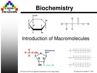
BIOCHEMISTRY ( BIO-100 ). Credit Hrs 4 (3-1). Course Contents. Introduction of biochemistry Biomolecules The Molecules and Chemical Reactions of Life Amino Acids and Proteins Simple and Complex Carbohydrates Lipids and Membranes Nucleotides and Nucleic Acids Vitamins and Cofactors

E N D
BIOCHEMISTRY (BIO-100) Credit Hrs 4 (3-1)
Course Contents • Introduction of biochemistry • Biomolecules • The Molecules and Chemical Reactions of Life • Amino Acids and Proteins • Simple and Complex Carbohydrates • Lipids and Membranes • Nucleotides and Nucleic Acids • Vitamins and Cofactors • Biochemical Reactions • Enzymes • Metabolic Pathways • Carbohydrate Metabolism • Lipid Metabolism • Amino Acid Metabolism • Molecular Genetics • DNA and RNA • Translation and the Genetic Code LAB WORK • Introduction of biochemistry lab and biosafety • Units of measurements • Buffer solution preparation • Determination of PH and POH • Numerical problems of PH and POH • Test on carbohydrates • Test on proteins • Test on lipids
Recommended Books • Fundamentals of Biochemistry: Life at the Molecular Level byVoet, Donald, Judith G. Voet, and Charlotte W. Pratt. • Concepts in biochemistry by Rodney Boyer. 3rd Edition. • Lippincott’s illustrated reviews: Lippincott Williams & Wilkins. • Biochemistry by Geoffrey Zubay McGraw-Hill. • Biochemistry by Donald Voet, Judith G. Voet 4, illustrated John Wiley & Sons. • Basic concepts in biochemistry: a student’s survival guide by Hiram F. Gilbert • Principles of biochemistry by Albert L. Lehninger, David L. Nelson, Michael M. Cox • Concepts in biochemistry by Rodney F. Boyer.
Introduction to Carbohydrates YasirWaheed
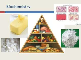
Carbohydrates are the most abundant organic molecules in nature. • They have a wide range of functions, including providing a significant fraction of the dietary calories for most organisms, acting as a storage form of energy in the body, and serving as cell membrane components that mediate some forms of intercellular communication. • Carbohydrates also serve as a structural component of many organisms, including the cell walls of bacteria, the exoskeleton of many insects, and the fibrous cellulose of plants. • The empiric formula for many of the simpler carbohydrates is (CH2O)n, hence the name “hydrate of carbon.”
CLASSIFICATION AND STRUCTURE OFCARBOHYDRATES • Monosaccharides (simple sugars) can be classified according to the number of carbon atoms they contain. Examples of some monosaccharidescommonly found in humans are listed in Figure 7.1. Figure 7.1 Examples of monosaccharides found in humans, classified according to the number of carbons they contain.
Carbohydrates with an aldehyde as their most oxidized functional group are called aldoses, whereas those with a keto as their most oxidized functional group are called ketoses (Figure 7.2). For example, glyceraldehyde is an aldose, whereas dihydroxy acetone is a ketose. Figure 7.2 Examples of an aldose (A) and a ketose(B) sugar.
Mono saccharides can be linked by glycosidic bonds to create larger structures (Figure 7.3). • Disaccharides contain two mono saccharide units, oligo saccharides contain from three to about ten monosaccharide units, whereas polysaccharides contain more than ten monosaccharide units, and can be hundreds of sugar units in length. Figure 7.3 A glycosidic bond between two hexoses producing a disaccharide.
Isomers and epimers • Compounds that have the same chemical formula but have different structures are called isomers. For example, fructose, glucose, mannose, and galactose are all isomers of each other, having the same chemical formula, C6H12O6. • Carbohydrate isomers that differ in configuration around only one specific carbon atom are defined as epimersof each other. For example, glucose and galactose are C-4 epimers—their structures differ only in the position of the –OH group at carbon 4. [Note: The carbons in sugars are numbered beginning at the end that contains the carbonyl carbon—that is, the aldehyde or keto group (Figure 7.4).] Glucose and mannose are C-2 epimers. • However, galactose and mannose are NOT epimers—they differ in the position of –OH groups at two carbons (2 and 4) and are, therefore, defined only as isomers (see Figure 7.4).
Joining of monosaccharides • Monosaccharides can be joined to form disaccharides, oligosaccharides, and polysaccharides. • Important disaccharides include lactose (galactose+ glucose), sucrose (glucose + fructose), and maltose (glucose + glucose). • Important polysaccharides include branched glycogen (from animal sources) and starch (plant sources) and unbranched cellulose (plant sources); each is a polymer of glucose. • The bonds that link sugars are called glycosidic bonds. These are formed by enzymes known as glycosyltransferases that use nucleotide sugars such as UDP-glucose as substrates. • Glycosidic bonds between sugars are named according to the numbers of the connected carbons, and with regard to the position of the anomeric hydroxyl group of the sugar involved in the bond. If this anomeric hydroxyl is in the α configuration, the linkage is an α-bond. If it is in the β configuration, the linkage is a β-bond. Lactose, for example, is synthesized by forming a glycosidic bond between carbon 1 of β-galactoseand carbon 4 of glucose. The linkage is, therefore, a β(1→4) glycosidic bond (see Figure 7.3).
Complex carbohydrates • Carbohydrates can be attached by glycosidic bonds to non-carbohydrate structures, including purine and pyrimidine bases (found in nucleic acids), aromatic rings (such as those found in steroids and bilirubin), proteins (found in glycoproteins and proteoglycans), and lipids (found in glycolipids). • 1. N- and O-glycosides: If the group on the non-carbohydrate molecule to which the sugar is attached is an –NH2 group, the structure is an N-glycoside and the bond is called an N-glycosidic link. If the group is an –OH, the structure is an O-glycoside, and the bond is an O-glycosidic link (Figure 7.7).
Figure 7.7 Glycosides: examples of N- and O-glycosidicbonds.
DIGESTION OF DIETARY CARBOHYDRATES • The principal sites of dietary carbohydrate digestion are the mouth and intestinal lumen. • This digestion is rapid and is catalyzed by enzymes known as glycoside hydrolases (glycosidases) that hydrolyze glycosidic bonds. • Because there is little monosaccharide present in diets of mixed animal and plant origin, the enzymes are primarily endoglycosidases that hydrolyze polysaccharides and oliosaccharides into disaccharidases and hydrolysetri- and disaccharides into their reducing sugar components (Figure 7.8). • Glycosidasesare usually specific for the structure and configuration of the glycosyl residue to be removed, as well as for the type of bond to be broken. The final products of carbohydrate digestion are the monosaccharides, glucose, galactose and fructose, which are absorbed by cells of the small intestine.
Digestion of Disaccharidases 1. Lactase hydrolyses lactose into two molecules, glucose and galactose: Lactase Lactose Glucose + Galactose 2. Maltase hydrolyses maltose into two molecules of glucose: Maltase Maltose Glucose + Glucose 3. Sucrasehydrolyses sucrose into two molecules of glucose and fructose: Sucrase Sucrose Glucose + Fructose 15
Figure 7.8 Hydrolysis of a glycosidic bond. Figure 7.9 Degradation of dietary glycogen by salivary or pancreatic α-amylase.
Abnormal degradation of disaccharides • The overall process of carbohydrate digestion and absorption is so efficient in healthy individuals that ordinarily all digestible dietary carbohydrate is absorbed by the time the ingested material reaches the lower jejunum.
Digestive enzyme deficiencies Genetic deficiencies of the individual disaccharidasesresult in disaccharide intolerance. Alterations in disaccharide degradation can also be caused by a variety of intestinal diseases, malnutrition, or drugs that injure the mucosa of the small intestine.
2. Lactose intolerance: More than three quarters of the world’s adults are lactose intolerant. The age-dependent loss of lactase activity represents a reduction in the amount of enzyme rather than a modified inactive enzyme. It is thought to be caused by small variations in the DNA sequence of a region on chromosome 2 that controls expression of the gene for lactase. Treatment for this disorder is to reduce consumption of milk.
3. Sucrase-isomaltase complex deficiency: This deficiency results in an intolerance of ingested sucrose. The disorder is found in about 10% of the people of Greenland and Canada, whereas 2% of North Americans are heterozygous for the deficiency. Treatment includes the dietary restriction of sucrose, and enzyme replacement therapy. 4. Diagnosis: Identification of a specific enzyme deficiency can be obtained by performing oral tolerance tests with the individual di - saccharides. Measurement of hydrogen gas in the breath is a reliable test for determining the amount of ingested carbohydrate not absorbed by the body, but which is metabolized instead by the intestinal flora (see Figure 7.11).
Figure 11-1. The relative permeability of a synthetic lipid bilayer to different classes of molecules. The smaller the molecule and, moreimportantly, the less strongly it associates with water, the more rapidly the molecule diffuses across the bilayer.
There Are Two Main Classes of Membrane Transport Proteins: Carriers and Channels • Carrier proteins (also called carriers, permeases, or transporters) bind the specific solute to be transported and undergo a series of conformational changes to transfer the bound solute across the membrane. • Channel proteins, in contrast, interact with the solute to be transported much more weakly. They form aqueous pores that extend across the lipid bilayer; when these pores are open, they allow specific solutes (usually inorganic ions of appropriate size and charge) to pass through them and thereby cross the membrane. Transport through channel proteins occurs at a much faster rate than transport mediated by carrier proteins.
Figure 11-3. Carrier proteins and channel proteins. (A) A carrier protein alternates between two conformations, so that the solute-binding site is sequentially accessible on one side of the bilayer and then on the other. (B) In contrast, a channel protein forms a water-filled pore across the bilayer through which specific solutes can diffuse.
Figure 11-4. Passive and active transport compared. (A) Passive transport down an electrochemical gradient occurs spontaneously, either by simple diffusion through the lipid bilayer or by facilitated diffusion through channels and passive carriers. By contrast, active transport requires an input of metabolic energy and is always mediated by carriers that harvest metabolic energy to pump the solute against its electrochemical gradient.
Carrier Proteins and Active Membrane Transport • Carrier protein has one or more specific binding sites for its solute (substrate). It transfers the solute across the lipid bilayer by undergoing reversible conformational changes that alternately expose the solute-binding site first on one side of the membrane and then on the other. • Coupled carriers couple the uphill transport of one solute across the membrane to the downhill transport of another. • 2. ATP-driven pumps couple uphill transport to the hydrolysis of ATP. • 3. Light-driven pumps, which are found mainly in bacterial cells, couple uphill transport to an input of energy from light, as with bacterio-rhodopsin
Figure 11-8. Three ways of driving active transport. The actively transported molecule is shown in yellow, and the energy source is shown in red.
Figure 11-9. Three types of carrier-mediated transport. This schematic diagram shows carrier proteins functioning as uniporters, symporters, and antiporters.
One way in which a glucose carrier can be driven by a Na+ gradient. The carrier oscillates between two alternate states, A and B. In the A state, the protein is open to the aextracellular space; in the B state, it is open to the cytosol. Binding of Na+ and glucose is cooperative that is, the binding of either ligand induces a conformational change that greatly increases the protein's affinity for the other ligand. Since the Na+ concentration is much higher in the extracellular space than in the cytosol, glucose is more likely to bind to the carrier in the A state. Therefore, both Na+ and glucose enter the cell (via an A to B transition) much more often than they leave it (via B to A transition). The overall result is the net transport of both Na+ and glucose into the cell. Because the binding is cooperative, if one of the two solutes is missing, the other fails to bind to the carrier. Thus, the carrier undergoes a conformational switch between the two states only if both solutes or neither are bound.