Download
1 / 74
740 likes | 758 Views
Explore different types of epithelial tissues and connective tissues through high-resolution microscope images. Learn to identify unique characteristics and structures.

E N D
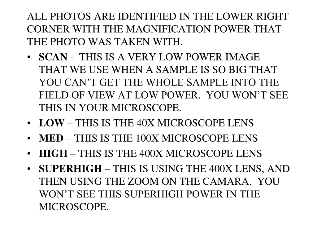
ALL PHOTOS ARE IDENTIFIED IN THE LOWER RIGHT CORNER WITH THE MAGNIFICATION POWER THAT THE PHOTO WAS TAKEN WITH. • SCAN - THIS IS A VERY LOW POWER IMAGE THAT WE USE WHEN A SAMPLE IS SO BIG THAT YOU CAN’T GET THE WHOLE SAMPLE INTO THE FIELD OF VIEW AT LOW POWER. YOU WON’T SEE THIS IN YOUR MICROSCOPE. • LOW – THIS IS THE 40X MICROSCOPE LENS • MED – THIS IS THE 100X MICROSCOPE LENS • HIGH – THIS IS THE 400X MICROSCOPE LENS • SUPERHIGH – THIS IS USING THE 400X LENS, AND THEN USING THE ZOOM ON THE CAMARA. YOU WON’T SEE THIS SUPERHIGH POWER IN THE MICROSCOPE.
EVERYTHING THAT YOU ARE RESPONSIBLE FOR ON THE HISTOLOGY IS LABELED AT LEAST ONCE IN THIS PPT. THERE ARE ALSO EXPLANATIONS OF WHAT MAKES SOMETHING DISTINCTIVE, SUCH THAT YOU CAN IDENTIFY IT.
ARTERY EPITHELIAL TISSUES ARE EITHER GLANDULAR OR LINING. HERE WE’RE LOOKING AT THE LINING AND WE’LL USE THE ARTERY LOW VEIN
LINING! MED
SEE HOW THE TEXTURE OF THE LINING IS DIFFERENT THAN THE UNDERLYING STUFF (MUSCLE). THAT IS THE SINGLE LAYER OF FLAT (SQUAMOUS) CELLS. HIGH
medulla This is a whole kidney, which is divided into layers. The middle layer is called the medulla, and is where we’ll find our simple cuboidal epithelium SCAN
SEE THAT THERE ARE DIFFERENT TEXTURES IN THIS SLIDE? THE NATURE OF THE KIDNEY IS THAT THESE TUBULES ALL SWEEP DOWN TOWARDS THE SAME PLACE TO EXIT THE KIDNEY. SO A SECTION THRU THE KIDNEY IS GOING TO CATCH SOME OF THE TUBULES IN CROSS SECTION AND SOME IN LONGITUDINAL SECTION. WE’LL SEE HIGH POWER OF BOTH AREAS LOW
THIS IS MEDIUM POWER OF THE LEFT PORTION OF THE PREVIOUS PHOTO – THE LONGITUDINAL SECTIONS… MED
Simple cuboidal epi lining - a cross section of a tubule HIGH
Simple cuboidal epi lining a longitudinal section of a tubule HIGH
WHY DO THESE CELLS APPARENTLY HAVE NO NUCLEI? Simple cuboidal epi lining a longitudinal section of a tubule HIGH
THE STOMACH HAS BOTH LINING EPITHELIUM AND GLANDY EPITHELIUM. THE VERY EDGE IS THE LINING AND ALL THE PURPLE STUFF UNDER IT ARE THE GASTRIC GLANDS LOW
SEE HOW THE LINING CELLS ARE TALL AND HAVE NUCLEI AT THE BASE, AND HOW THE NUCLEI ARE LINED UP IN A NICE ROW? HIGH
Note the dark staining at the bottom of the tissue, reflecting the packed nuclei. LOW
As the cells are pushed away from the basal layer, they get flatter, and the nuclei get spread out too. MED
flattening HIGH
In this slide the flattening isn’t as obvious, but we can still see the the density of the nuclei decrease as the cells are pushed to the surface HIGH
THIS IS ALL ADIPOSE TISSUE Look here for the epi LOW
The basal layer of the st. sq epi curves around as the epidermis invaginates into the dermis. There are clearly layers here – we’ll get to them later. For now just note the concentrated, darkly staining nuclei at the base, thinning out a bit as they get pushed off the basal layer. Also note the thick superficial layer of pure keratin. MED
KERATIN BORDER BETWEEN THE EPI AND THE UNDERLYING TISSUE THINNING AND FLATTENING HIGH
LINING THE LUMEN… BLOOD VESSELS JEJUNUM LOW A SNIPPET OF THE PANCREAS
THIS IS A SINGLE LAYER, SO LOOK AT HOW THE NUCLEI ARE LINED UP, AND LOOK AT THE EDGE OF THE CELLS, THAT DARKEaNED LINE ARE THE PACKED MICROVILLI (TOO SMALL TO BE SEEN ONE BY ONE, BUT SO DENSE THEY COLLECTIVELUY STAIN DARKER HIGH
WE CAN SEE THE CILIA THIS IS A BLOB OF LYMPHATIC TISSUE - WBCS THESE ARE COLUMNAR CELLS, BUT THE NUCLEI AREN’T IN A NICE ROW – THEY’RE IN A JUMBLED MESS HIGH THIS IS MUSCUS GUNK ON THE SURFACE OF THE CILIA
Bladder slides are tough to make, the layer under the epithelial layer separates as the section is made, resulting in all those spaces, which are ‘artifacts’ MED
Transitional epi is designed to be able to stretch and collapse as the bladder slowly fills and then quickly collapses. The nuclei are jumbled, but are clearly NOT columnar. HIGH
WHEREAS EPI CONSISTS OF TIGHTLY PACKED CELLS WITH ALMOST NO MATERIAL BETWEEN THEM, CT IS MOSTLY ‘INSTITIAL’ STUFF (FIBERS AND SUCH), AND THE CELLS ARE WIDELY SCATTERED. EVERYWHERE YOU SEE A NUCLEUS IS A CELL LOW
Look at how scattered the nuclei (and therefore the cells) are. Most of the mass of CT is extracellular material, made by the fibroblasts. All the pinkish and thin black strands are the fibers that give the tissue (along with the nature of the ground substance) its character This is loose ct, disorganized with the fibers not nearly as compacted as Dense CT HIGH
Back to the skin to see DICT. It’s here, below the epithelial layer, but above the fatty layer LOW
There are a bunch of accessory structures here. Ignore them for now. Look at the disorganized stuff that is the ‘stuff ‘ of this layer MED
Ignore all the extra stuff in the layer – there are blood vessles and glands here – and look at the thick pinkish collagen fibers. They’re not regularly arranged and there are way more of them than in LCT HIGH
EVEN AT LOW POWER WE CAN SEE HOW THERE IS AN ORGANIZATION, REFLECTING THE FIBERS BEING PARALLEL TO EACH OTHER AND GIVING THE TISSUE POINT-TO-POINT STRENGTH AS OPPOSED TO DISTRIBUTED STRENGTH LOW