Download
1 / 78
780 likes | 803 Views
This overview discusses the role of hormones and other signaling molecules in the body, including their release, target receptors, and cellular response pathways. It explores the different types of signaling molecules, such as hormones, local regulators, neurotransmitters, neurohormones, and pheromones. The classification and characteristics of hormones, including their chemical classes and cellular response pathways, are also explained.

E N D
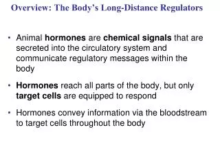
Overview: The Body’s Long-Distance Regulators • Animal hormones are chemical signals that are secreted into the circulatory system and communicate regulatory messages within the body • Hormones reach all parts of the body, but only target cells are equipped to respond (have the right receptor molecules) • Insect metamorphosis is regulated by hormones
Two systems coordinate communication throughout the body: the endocrine system and the nervous system • The endocrine system secretes hormones that coordinate slower but longer-acting responses including reproduction, development, energy metabolism, growth, and behavior • The nervous system conveys high-speed electrical signals along specialized cells called neurons; these signals regulate other cells
Concept 1: Hormones and other signaling molecules bind to target receptors, triggering specific response pathways • Chemical signals bind to receptor proteins on target cells • Only target cells respond to the signal
Types of Secreted Signaling Molecules • Secreted chemical signals include • Hormones • Local regulators • Neurotransmitters • Neurohormones • Pheromones
Hormones • Endocrine signals (hormones) are secreted into extracellular fluids and travel via the bloodstream • Endocrine glands are ductless and secrete hormones directly into surrounding fluid
Fig. 45-2 • Endocrine signaling • Blood • vessel • Response
Exocrine glands have ducts and secrete substances onto body surfaces or into body cavities (for example, tear ducts)
Neurotransmitters and Neurohormones • Neurons (nerve cells) contact target cells at synapses • At synapses, neurons often secrete chemical signals called neurotransmitters that diffuse a short distance to bind to receptors on the target cell • Neurotransmitters play a role in sensation, memory, cognition, and movement
Fig. 45-2b • Synapse • Neuron • Response • (d) Synaptic signaling • Neurosecretory • cell • Blood • vessel • Response • (e) Neuroendocrine signaling
Neurohormones are a class of hormones that originate from neurons in the brain and diffuse through the bloodstream
Pheromones • Pheromones are chemical signals that are released from the body and used to communicate with other individuals in the species • Pheromones mark trails to food sources, warn of predators, and attract potential mates
Chemical Classes of Hormones • Three major classes of molecules function as hormones in vertebrates: • Polypeptides (proteins and peptides) • Amines derived from amino acids • Steroid hormones
Lipid-soluble hormones (steroid hormones) pass easily through cell membranes, while water-soluble hormones (polypeptides and amines) do not • The solubility of a hormone correlates with the location of receptors inside or on the surface of target cells
Fig. 45-3 • Water-soluble • Lipid-soluble • 0.8 nm • Polypeptide: • Insulin • Steroid: • Cortisol • Amine: • Epinephrine • Amine: • Thyroxine
Cellular Response Pathways • Water and lipid soluble hormones differ in their paths through a body • Water-soluble hormones are secreted by exocytosis, travel freely in the bloodstream, and bind to cell-surface receptors • Lipid-soluble hormones diffuse across cell membranes, travel in the bloodstream bound to transport proteins, and diffuse through the membrane of target cells
Signaling by any of these hormones involves three key events: • Reception • Signal transduction • Response
Fig. 45-5-1 • Fat-soluble • hormone • Water- • soluble • hormone • Transport • protein • Signal receptor • TARGET • CELL • Signal • receptor • NUCLEUS • (a) • (b)
Fig. 45-5-2 • Fat-soluble • hormone • Water- • soluble • hormone • Transport • protein • Signal receptor • TARGET • CELL • OR • Signal • receptor • Cytoplasmic • response • Gene • regulation • Cytoplasmic • response • Gene • regulation • NUCLEUS • (a) • (b)
Pathway for Water-Soluble Hormones • Binding of a hormone to its receptor initiates a signal transduction pathway leading to responses in the cytoplasm, enzyme activation, or a change in gene expression Animation: Water-Soluble Hormone
The hormone epinephrine (formerly known as adrenalin) has multiple effects in mediating the body’s response to short-term stress • Epinephrine binds to receptors on the plasma membrane of liver cells • This triggers the release of messenger molecules that activate enzymes and result in the release of glucose into the bloodstream
Fig. 45-6-1 • Epinephrine • Adenylyl • cyclase • G protein • GTP • G protein-coupled • receptor • ATP • Second • messenger • cAMP
Fig. 45-6-2 • Epinephrine • Adenylyl • cyclase • G protein • GTP • G protein-coupled • receptor • ATP • Second • messenger • cAMP • Protein • kinase A • Inhibition of • glycogen synthesis • Promotion of • glycogen breakdown
Pathway for Lipid-Soluble Hormones • The response to a lipid-soluble hormone is usually a change in gene expression • Steroids, thyroid hormones, and the hormonal form of vitamin D enter target cells and bind to protein receptors in the cytoplasm or nucleus • Protein-receptor complexes then act as transcription factors in the nucleus, regulating transcription of specific genes Animation: Lipid-Soluble Hormone
Fig. 45-7-1 • Hormone • (estradiol) • Estradiol • (estrogen) • receptor • Plasma • membrane • Hormone-receptor • complex
Fig. 45-7-2 • Hormone • (estradiol) • Estradiol • (estrogen) • receptor • Plasma • membrane • Hormone-receptor • complex • DNA • Protein • mRNA
Multiple Effects of Hormones • The same hormone may have different effects on target cells that have • Different receptors for the hormone • Different signal transduction pathways • Different proteins for carrying out the response • A hormone can also have different effects in different species
Fig. 45-8-1 • Same receptors but different • intracellular proteins (not shown) • Epinephrine • Epinephrine • receptor • receptor • Glycogen • deposits • Vessel • dilates. • Glycogen • breaks down • and glucose • is released. • (a) Liver cell • (b) Skeletal muscle • blood vessel
Fig. 45-8-2 • Same receptors but different • intracellular proteins (not shown) • Different receptors • Epinephrine • Epinephrine • Epinephrine • Epinephrine • receptor • receptor • receptor • receptor • Glycogen • deposits • Vessel • dilates. • Vessel • constricts. • Glycogen • breaks down • and glucose • is released. • (a) Liver cell • (c) Intestinal blood • vessel • (b) Skeletal muscle • blood vessel
Concept 2: Negative feedback and antagonistic hormone pairs are common features of the endocrine system • Hormones are assembled into regulatory pathways
Fig. 45-10 • Major endocrine glands: • Hypothalamus • Pineal gland • Pituitary gland • Organs containing • endocrine cells: • Thyroid gland • Thymus • Parathyroid glands • Heart • Liver • Adrenal • glands • Stomach • Pancreas • Kidney • Testes • Small • intestine • Kidney • Ovaries
Simple Hormone Pathways • Hormones are released from an endocrine cell, travel through the bloodstream, and interact with the receptor or a target cell to cause a physiological response
A negative feedback loop inhibits a response by reducing the initial stimulus • Negative feedback regulates many hormonal pathways involved in homeostasis
Insulin and Glucagon: Control of Blood Glucose • Insulinand glucagonare antagonistic (have opposite effects) hormones that help maintain glucose homeostasis • The pancreashas clusters of endocrine cells called islets of Langerhans with alpha cells that produce glucagon and beta cells that produce insulin
Fig. 45-12-1 • Insulin • Beta cells of • pancreas • release insulin • into the blood. • STIMULUS: • Blood glucose level • rises. • Homeostasis: • Blood glucose level • (about 90 mg/100 mL)
Fig. 45-12-2 • Body cells • take up more • glucose. • Insulin • Beta cells of • pancreas • release insulin • into the blood. • Liver takes • up glucose • and stores it • as glycogen. • STIMULUS: • Blood glucose level • rises. • Blood glucose • level declines. • Homeostasis: • Blood glucose level • (about 90 mg/100 mL)
Fig. 45-12-3 • Homeostasis: • Blood glucose level • (about 90 mg/100 mL) • STIMULUS: • Blood glucose level • falls. • Alpha cells of pancreas • release glucagon. • Glucagon
Fig. 45-12-4 • Homeostasis: • Blood glucose level • (about 90 mg/100 mL) • STIMULUS: • Blood glucose level • falls. • Blood glucose • level rises. • Alpha cells of pancreas • release glucagon. • Liver breaks • down glycogen • and releases • glucose. • Glucagon
Fig. 45-12-5 • Body cells • take up more • glucose. • Insulin • Beta cells of • pancreas • release insulin • into the blood. • Liver takes • up glucose • and stores it • as glycogen. • STIMULUS: • Blood glucose level • rises. • Blood glucose • level declines. • Homeostasis: • Blood glucose level • (about 90 mg/100 mL) • STIMULUS: • Blood glucose level • falls. • Blood glucose • level rises. • Alpha cells of pancreas • release glucagon. • Liverbreaks • downglycogen • andreleases • glucose. • Glucagon
Target Tissues for Insulin and Glucagon • Insulin reduces blood glucose levels by • Promoting the cellular uptake of glucose • Slowing glycogen breakdown in the liver • Promoting fat storage
Glucagon increases blood glucose levels by • Stimulating conversion of glycogen to glucose in the liver • Stimulating breakdown of fat and protein into glucose
Diabetes Mellitus • Diabetes mellitus is perhaps the best-known endocrine disorder • It is caused by a deficiency of insulin or a decreased response to insulin in target tissues • It is marked by abnormally high blood glucose levels
Type I diabetes mellitus (insulin-dependent) is an autoimmune disorder in which the immune system destroys pancreatic beta cells • Type II diabetes mellitus (non-insulin-dependent) involves insulin deficiency or reduced response of target cells due to change in insulin receptors
Concept 3: The endocrine and nervous systems act individually and together in regulating animal physiology • Signals from the nervous system initiate and regulate endocrine signals
Coordination of Endocrine and Nervous Systems in Invertebrates • In insects, molting and development are controlled by a combination of hormones: • A brain hormone stimulates release of ecdysone from the prothoracic glands • Juvenile hormone promotes retention of larval characteristics • Ecdysone promotes molting (in the presence of juvenile hormone) and development (in the absence of juvenile hormone) of adult characteristics
Fig. 45-13-1 • Brain • Neurosecretory cells • Corpus cardiacum • PTTH • Corpus allatum • Prothoracic • gland • Ecdysone • Juvenile • hormone • (JH) • EARLYLARVA
Fig. 45-13-2 • Brain • Neurosecretory cells • Corpus cardiacum • PTTH • Corpus allatum • Prothoracic • gland • Ecdysone • Juvenile • hormone • (JH) • EARLYLARVA • LATER • LARVA
Fig. 45-13-3 • Brain • Neurosecretory cells • Corpus cardiacum • PTTH • Corpus allatum • Low • JH • Prothoracic • gland • Ecdysone • Juvenile • hormone • (JH) • EARLYLARVA • LATER • LARVA • PUPA • ADULT