Download
1 / 40
400 likes | 489 Views
Explore the intricacies of DNA replication, mutation types, repair mechanisms, and cellular processes involved in maintaining genetic integrity. Learn about the semi-conservative nature of DNA replication, various mutations, and repair pathways.

E N D
3. DNA Replication, Mutation, Repair a). DNA replication i). Cell cycle/ semi-conservative replication ii). Initiation of DNA replication iii). Discontinuous DNA synthesis iv). Components of the replication apparatus b). Mutation i). Types and rates of mutation ii). Spontaneous mutations in DNA replication iii). Lesions caused by mutagens c). DNA repair i). Types of lesions that require repair ii). Mechanisms of repair Proofreading by DNA polymerase Mismatched repair Excision repair iii). Defects in DNA repair or replication
Themammalian cell cycle DNA synthesis and histone synthesis Rapid growth and preparation for DNA synthesis S phase G0 G1 phase Quiescent cells G2 phase Growth and preparation for cell division M phase Mitosis
DNA replication is semi-conservative Parental DNA strands Each of the parental strands serves as a template for a daughter strand Daughter DNA strands
Origins of DNA replication on mammalian chromosomes origins of DNA replication (every ~150 kb) 5’ 3’ 3’ 5’ bidirectional replication replication bubble fusion of bubbles 5’ 3’ 3’ 5’ daughter chromosomes 5’ 3’ 3’ 5’
5’ 3’ 3’ 5’ A A A A A A A A A dnaB and dnaC proteins bind to the single-stranded DNA A A A B C A A A dnaB further unwinds the helix Initiation of DNA synthesis at the E. coli origin (ori) origin DNA sequence binding of dnaA proteins DNA melting induced by the dnaA proteins dnaA proteins coalesce
A A A C A A A A A A C A A A dnaG (primase) binds... G B dnaB further unwinds the helix and displaces dnaA proteins ...and synthesizes an RNA primer G B RNA primer
G B C Primasome dna B (helicase) dna C dna G (primase) template strand 5’ 3’ OH 3’ 5’ RNA primer (~5 nucleotides)
DNA polymerase 5’ 3’ 5’ RNA primer 3’ 5’ newly synthesized DNA
DNA DNA • Reaction catalyzed by DNA polymerase • all DNA polymerases require a primer with afree 3’ OH group • all DNA polymerases catalyze chain growth in a 5’ to 3’ direction • some DNA polymerases have a 3’ to 5’ proofreading activity
Discontinuous synthesis of DNA 5’ 3’ 5’ 3’ 3’ 5’ 5’ 3’ 3’ 5’ 3’ 5’ Because DNA is always synthesized in a 5’ to 3’ direction, synthesis of one of the strands... 5’ 3’ ...has to be discontinuous. This is the lagging strand.
Each replication fork has a leading and a lagging strand leading strand (synthesized continuously) replication fork replication fork 5’ 3’ 5’ 3’ 3’ 5’ 5’ 3’ 3’ 5’ 3’ 5’ lagging strand (synthesized discontinuously) • The leading and lagging strand arrows show the direction • of DNA chain elongation in a 5’ to 3’ direction • The small DNA pieces on the lagging strand are called • Okazaki fragments (100-1000 bases in length)
RNA primer direction of leading strand synthesis 3’ 5’ replication fork 5’ 3’ 3’ 5’ direction of lagging strand synthesis
Movement of the replication fork 5’ 3’ 5’ 3’
Movement of the replication fork 5’ RNA primer Okazaki fragment RNA primer
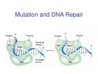
RNA primer pol III 5’ 5’ 3’ DNA polymerase III initiates at the primer and elongates DNA up to the next RNA primer 5’ 5’ 3’ newly synthesized DNA (100-1000 bases) (Okazaki fragment) pol I 5’ 3’ DNA polymerase I inititates at the end of the Okazaki fragment and further elongates the DNA chain while simultaneously removing the RNA primer with its 5’ to 3’ exonuclease activity
newly synthesized DNA (Okazaki fragment) 5’ 3’ DNA ligase seals the gap by catalyzing the formation of a 3’, 5’-phosphodiester bond in an ATP-dependent reaction 5’ 3’
G DNA ligase C B pol I Proteins at the replication fork in E. coli Rep protein (helicase) 3’ 5’ pol III 5’ 3’ Primasome Single-strand binding protein (SSB) pol III DNA gyrase - this is a topoisomerase II, which breaks and reseals the DNA to introduce negative supercoils ahead of the fork
Components of the replication apparatus dnaA binds to origin DNA sequence Primasome dnaB helicase (unwinds DNA at origin) dnaC binds dnaB dnaG primase (synthesizes RNA primer) DNA gyrase introduces negative supercoils ahead of the replication fork Rep protein helicase (unwinds DNA at fork) SSB binds to single-stranded DNA DNA pol III primary replicating polymerase DNA pol I removes primer and fills gap DNA ligase seals gap by forming 3’, 5’-phosphodiester bond
Properties of DNA polymerases DNA polymerases of E. coli_ pol I pol II pol III (core) Polymerization: 5’ to 3’ yes yes yes Proofreading exonuclease: 3’ to 5’ yes yes yes Repair exonuclease: 5’ to 3’ yes no no DNA polymerase III is the main replicating enzyme DNA polymerase I has a role in replication to fill gaps and excise primers on the lagging strand, and it is also a repair enzyme • all DNA polymerases require a primer with a free 3’ OH group • all DNA polymerases catalyze chain growth in a 5’ to 3’ direction • some DNA polymerases have a 3’ to 5’ proofreading activity
Properties of DNA polymerases DNA polymerases of humans a b g d e Location nucl nucl mito nucl nucl Replication yes no yes yes yes Repair no yes no yes yes3 Functions 5’ to 3’ polymerase yes yes yes yes yes 3’ to 5’ exonuclease no no yes yes yes 5’ to 3’ exonuclease1 no no no no no Primase yes no no no no Associates with PCNA2 no no no yes yes Processivity low high Strand synthesis lagging* repair both leading* lagging* * see notes below 1 activity present in associated proteins 2 Proliferating Cell Nuclear Antigen – “sliding clamp” 3 involved in transcription-linked DNA repair
pol d DNA ligase Proteins at the replication fork in humans leading strand helicase 3’ 5’ PCNA 5’ 3’ 5’ to 3’ exo associated with the complex SSB pol a (or pol d) pol e topoisomerases I and II primase activity associated with pol a lagging strand
Mutation Types and rates of mutation Type Mechanism Frequency________ Genome chromosome 10-2 per cell division mutation missegregation (e.g., aneuploidy) Chromosome chromosome 6 X 10⁻⁴ per cell division mutation rearrangement (e.g., translocation) Gene base pair mutation 10-10 per base pair per mutation (e.g., point mutation, cell division or or small deletion or 10-5 - 10-6 per locus per insertion generation
Mutation rates* of selected genes Gene New mutations per 106 gametes Achondroplasia 6 to 40 Aniridia 2.5 to 5 Duchenne muscular dystrophy 43 to 105 Hemophilia A 32 to 57 Hemophilia B 2 to 3 Neurofibromatosis -1 44 to 100 Polycystic kidney disease 60 to 120 Retinoblastoma 5 to 12 *mutation rates (mutations / locus / generation) can vary from 10-4 to 10-7 depending on gene size and whether there are “hot spots” for mutation (the frequency at most loci is 10-5 to 10-6).
Polymorphisms exist in the genome • the number of existing polymorphisms is ~1 per 500 bp • there are ~5.8 million differences per haploid genome • polymorphisms were caused by mutations • New germline mutations • each sperm contains ~100 new mutations • a normal ejaculate has ~100 million sperm • 100 X 100 million = 10 billion new mutations • ~1 in 10 sperm carries a new deleterious mutation • at a rate of production of ~8 X 10⁷ sperm per day, • a male will produce a sperm with a new mutation • in the Duchenne muscular dystrophygene • approximately every 10 seconds.
Types of base pair mutations normal sequence CATTCACCTGTACCA GTAAGTGGACATGGT transition (T-A to C-G) transversion (T-A to G-C) CATCCACCTGTACCA GTAGGTGGACATGGT CATGCACCTGTACCA GTACGTGGACATGGT base pair substitutions transition: pyrimidine to pyrimidine transversion: pyrimidine to purine deletion insertion CATCACCTGTACCA GTAGTGGACATGGT CATGTCACCTGTACCA GTACAGTGGACATGGT deletions and insertions can involve one or more base pairs
Spontaneous mutations can be caused by tautomers Tautomeric forms of the DNA bases Adenine Cytosine AMINO IMINO
Tautomeric forms of the DNA bases Guanine Thymine KETO ENOL
Mutation caused by tautomer of cytosine Cytosine Normal tautomeric form Guanine Cytosine Rare imino tautomeric form Adenine • cytosine mispairs with adenine resulting in a transition mutation
C G and Mutation is perpetuated by replication C G C G C G and • replication of C-G should give daughter strands each with C-G C G C A • tautomer formation Cduring replication will result in mispairing • and insertion of an improper A in one of the daughter strands C A T A • which could result in a C-G to T-A transition mutation in the next • round of replication, or if improperly repaired
Chemical mutagens Deamination by nitrous acid
Derivation by hydroxylamine Alkylation by dimethyl sulfate causes depurination The formation of a quarternary nitrogen destabilizes the deoxyriboside bond and the base is released from deoxyribose
Summary of DNA lesions Missing base Acid and heat depurination (~104 purines per day per cell in humans) Altered base Ionizing radiation; alkylating agents Incorrect base Spontaneous deaminations cytosine to uracil adenine to hypoxanthine Deletion-insertion Intercalating reagents (acridines) Dimer formation UV irradiation Strand breaks Ionizing radiation; chemicals (bleomycin) Interstrand cross-links Psoralen derivatives; mitomycin C (Tautomer formation Spontaneous and transient)
Mechanisms of Repair • Mutations that occur during DNA replication are repaired when • possible by proofreading by the DNA polymerases • Mutations that are not repaired by proofreading are repaired • by mismatched (post-replication) repair followed by • excision repair • Mutations that occur spontaneously any time are repaired by • excision repair (base excision or nucleotide excision)
CH3 CH3 CH3 CH3 Mismatched (post-replication) repair • the parental DNA strands are • methylated on certain • adenine bases • mutations on the newly • replicated strand are • identified by scanning • for mismatches prior to • methylation of the newly • replicated DNA 5’ 3’ • the mutations are repaired • by excision repair mechanisms • after repair, the newly • replicated strand is methylated
Excision repair (base or nucleotide) deamination ATGCUGCATTGA TACGGCGTAACT uracil DNA glycosylase ATGCGCATTGA TACGGCGTAACT repair nucleases AT GCATTGA TACGGCGTAACT DNA polymerase b ATGCCGCATTGA TACGGCGTAACT DNA ligase ATGCCGCATTGA TACGGCGTAACT thymine dimer ATGCUGCATTGATAG TACGGCGTAACTATC excinuclease AT AG TACGGCGTAACTATC (~30 nucleotides) DNA polymerase b ATGCCGCATTGATAG TACGGCGTAACTATC DNA ligase ATGCCGCATTGATAG TACGGCGTAACTATC Base excision repair Nucleotide excision repair
Deamination of cytosine can be repaired Deamination of 5-methylcytosine cannot be repaired More than 30% of all single base changes that have been detected as a cause of genetic disease have occurred at 5’-mCG-3’ sites
Defects in DNA repair or replication • Xeroderma pigmentosum • Ataxia telangiectasia • Fanconi anemia • Bloom syndrome • Cockayne syndrome 100 human elephant cow Life span 10 hamster Correlation between DNA repair activity in fibroblast cells from various mammalian species and the life span of the organism rat mouse shrew 1 DNA repair activity
Defects in DNA repair or replication • All are associated with a high frequency of chromosome • and gene (base pair) mutations; most are also associated with a • predisposition to cancer, particularly leukemia • Xeroderma pigmentosum • caused by mutations in genes involved in nucleotide excision repair • associated with a 2000-fold increase of sunlight-induced • skin cancer and with other types of cancer such as melanoma • Ataxia telangiectasia • caused by gene that detects DNA damage • increased risk of X-ray • associated with increased breast cancer in carriers • Fanconi anemia • increased risk of X-ray • sensitivity to sunlight • Bloom syndrome • caused by mutations in a a DNA helicase gene • increased risk of X-ray • sensitivity to sunlight • Cockayne syndrome • caused by a defect in transcription-linked DNA repair • sensitivity to sunlight • Werner’s syndrome • caused by mutations in a DNA helicase gene • premature aging