Download
1 / 23
250 likes | 543 Views
Learn about primary and secondary injuries, muscle strains, joint sprains, and their classifications. Discover the symptoms, treatments, and complications of various muscle and joint traumas.

E N D
Mechanism of Injuries Primary Injury – An injury that results directly from trauma Extrinsic – external, something hitting the body Intrinsic – Results from stressors in the body Secondary Injury – Arise from an injury not being treated properly or return to participation to soon Chronic weakness Swelling Pain Inflammation Arthritis
Skin Trauma Wound Classifications • Blister – continuous rubbing causes fluid buildup below epidermis (Epidermis/Dermis) • Abrasion – Skin scrapped off • Skin Bruise (Contusion) – Compression or crushing force produces bleeding under the skin • Laceration – Wound in which skin is irregularly torn • Skin Avulsion – Skin completely ripped away • Puncture Wound – Penetration of skin by a sharp object
Muscle Trauma Muscle Trauma – Caused by three mechanisms • Compression Force • Tension Force • Shearing Force
Acute Muscle Injuries Acute Injury – Sudden Onset Contusion (bruise) – Caused by a blow to the muscle tissue Signs/Symptoms • Athlete reports being struck • Pain • Temporary paralysis • Palpation reveals hard, swollen area • Ecchymosis – Tissue discoloration (black & blue)
Three Classifications of Muscle Contusions 1st Degree Muscle Contusion • Little to no loss of movement • Minor pain on palpation • No loss of strength 2nd Degree Muscle Contusion • Moderate Pain • Ecchymosis • Swelling, hardness in muscle • Some loss of range of motion 3rd Degree Muscle Contusion • Severe pain & Swelling • Ecchymosis • Complete restriction of movement
Muscle Strain Muscle Strain – Stretch – Rip – Tear in muscle fibers or tendon, generally cause by abnormal muscle contraction 1st Degree Muscle Strain • Mild local pain , increases with muscle use • Minor loss of strength • Mild Swelling • Mild Ecchymosis • Local Tenderness 2nd Degree Muscle Strain – Moderate symptoms as above 3rd Degree Muscle Strain – Severe Injury, generally complete tear of muscle • Severe Pain • Complete Loss of Function • Palpable defect
Most Common Muscle Strains • Hamstring • Gastrocnemius • Quadriceps • Illiopsoas • Hips Adductors • Spinals • Deltoid • Rotator Cuff
Tendon Strain Tendon – 2X as strong as muscle, so single event usually occurs at belly of muscle, muscle/tendon junction or bony attachment Tendon Strain – usually caused by continually abnormal tension • Increased Collagen Tissue • Weakness in tendon
Muscle Cramp & Spasm Muscle Cramp – Spasm:Involuntary contraction of muscle • Clonic – MuscleTwitch – Contract/relax • Tonic – Rigid Muscle – no relaxation Muscle Cramp – • Electrolyte Imbalance • Fatigue Muscle Spasm – • Reflex reaction of muscle caused by direct trauma
Chronic Muscle Injuries Chronic Muscle Injury – Slow progressive injuries over a period of time • Repeated Acute Injuries • Constant Irritation by Poor Technique • Constant Stress beyond Physiological Limits • Acute Injuries improperly managed
Inflammation • Bursitis – Inflammation of Bursae • Pain on/over bursae • Swelling/ egg shape • Loss of function • Myositis Ossificans – Calcification within Muscle • Repeated trauma causes calcium deposits to form • Hard bumps in bed of muscle • Discovered on x-ray • Usual Sites are Biceps & Quadriceps
Complications to Muscle/tendon Injuries Atrophy – Shrinking/wasting away of muscle Contracture – Abnormal shortening of muscle with great resistance to stretch
Joint Injuries Synovial Joints – Freely moving joints Characteristics of a Synovial Joint • Synovial Capsule – covering encapsulating each joint • Ligaments - Intrinsic: part of capsule, Extrinsic: Outside of joint, not part of capsule. Support Joint • Synovial Fluid – Joint lubrication • Articular Cartilage – Cushions Bone Ends, provide no support
Joint Injuries Sprains – Twist, stretch, tearing of connective tissue – ligaments, cartilage, capsule. 1st Degree Sprain • Minimal pain • Minimal loss of function • Mild point tenderness • Little to no swelling • No instability 2nd Degree Sprain – moderate symptoms with slight instability 3rd Degree Sprain • Very painful • Major Loss of Function • Marked Instability • Severe Tenderness & Swelling
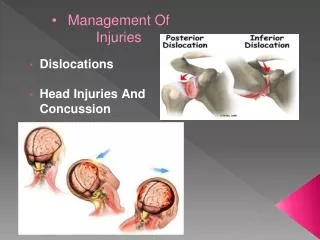
Joint Injuries Acute Synovitis – Inflammation of capsule caused by contusion or sprain • Pain on joint movement • Swelling • Skin sensitivity at joint capsule • If properly managed – heals quickly in a few days Subluxation – Dislocation • Subluxation – partial dislocation – moved but did not come out of joint • Dislocation – Total disunion of bone ends • Recognizing a Dislocation • Loss of function • Deformity • Swelling & point tenderness 1st Time Dislocation – Severe trauma, tearing, breaking, chipping of bone. Always treat 1st time dislocation like a fracture – Splint & Medical referral
Bone Trauma • Periostitis • Acute Fracture • Stress Fracture • Epiphyseal Damage Periostitis – Inflammation of periosteum (covering of bone) Bone Contusion/Bruise
Bone Trauma Acute Bone Fracture – Fracture vs. Break • Closed Fracture • Open Fracture (Compound Fracture) • Avulsion Fracture – pulled away piece of bone – ligament or tendon, chip • Epiphyseal Fracture – Injury to growth area – ages 10-16 yrs Stress Fracture – Exact etiology unknown • Overload stress caused by muscle • Altered stress caused by muscle fatigue • Rhythmic muscle vibration over long period of time • Vibration caused by ground reaction • Bones are more vulnerable to stress fracture the 1st few weeks of physical training
Causes of Stress Fractures • Too Much too Soon • Going From One Sport to Another Without Proper Training • Going Back to competition too soon after an Injury • Changing environment; running surfaces, gym to track, grass to concrete, shoes
Signs of Stress Fracture Shin Splints – Stress fracture • Swelling • Shin Splints – pain on initial activity & decreases once warm only to increase over time • Night Pain • Tenderness-Pain • Shin Splints more generalized pain • Stress Fracture more local (size of a quarter) pain on vibration • Most Common Sights of Stress Fractures • Tibia, Fibula, Metatarsal, Calcaneus, femur, Lumbar Spine, Ribs, Humerus
Nerve Trauma Nerve Trauma – caused by direct blow, stretch or swelling • Hypoesthesia – Diminished sense of feeling • Hyperesthesia – Increased feeling to pain or touch • Paraesthesia – Numbness, prickling, tingling • Referred Pain – Pain that is felt at another location other than actual site of original injury