Download
1 / 39
410 likes | 597 Views
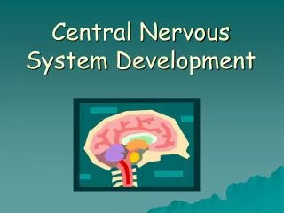
Dr. Ágoston Szél. Development of the Nervous System. Semmelweis University Department of Anatomy , Histology and Embryology 2018. ventricles. lateral ventricles. 3rd ventricle. cerebral aqueduct. 4th ventricle. central canal. Development of Brain Vesicles. secondary brain vesicles.

E N D
Dr. Ágoston Szél Development of the Nervous System Semmelweis University Department of Anatomy, Histology and Embryology 2018
ventricles lateral ventricles 3rd ventricle cerebral aqueduct 4th ventricle central canal Development of Brain Vesicles secondary brain vesicles primary brain vesicles end-brain (telencephalon) between-brain (diencephalon) forebrain (prosencephalon) midbrain (mesencephalon) midbrain (mesencephalon) behind-brain(metencephalon) pons, cerebellum hindbrain (rhombencephalon) medulla oblongata medulla brain(myelencephalon) spinal cord (medulla spinalis) spinal cord (medulla spinalis)
Development of Brain Vesicles rhombencephalic isthmus cephalic flexure cervical flexure pontine flexure mesodiencephalic groove telodiencephalic groove cephalic flexure cervical flexure pontine flexure
Transitional Segmentation of the Neural Tube (Neuromeres) isthmic neuromer rhombomers i r1 M mesencephalic neuromer r2 r3 P6 r4 r5 P5 r6 P4 r7-8 P3 P2 prosomeres P1 D 21
Drosophila embryo HomeoboxGenes Drosophila genes Chr 3 mouse embryo Chr 2 Chr 6 mouse genes Chr11 Chr 15
Coordinates of the Developing Neural Tube BMP, dorsalin D A P hox4 hox1 retinoic BMP acid V Sonic hedgehog
MigratoryPathways and NeurogeneDerivatives of theTruncalNeuralCrest neurogenin dorsal root, cranial nerve sensory ganglions ventrolateral pathway endothelin-3 neural crest pigment cells (melanocytes) dorsolateral pathway parasympathic andsympathic ganglions mesoderm of somites developing adrenal gland (medulla) glucocorticoid hormons parasympathic (submucous) plexus in the gut ventral pathway enterochromaffine cells Mash 1 (HLH)
MigratoryPathways and NeurogeneDerivatives of theTruncalNeuralCrest ventrolateral pathway neural crest Schwann’s cells satellite cells unipolar sensory neurons multipolar ganglion cells in sympathetic ganglions FGF, NGF chromaffine cells in adrenal glands glomus caroticum, parafollicular (C) cells prevertebral plexus
3. rhombomere Outmigration of NeuralCrest 2. rhombomere 1. rhombomere neurogene crest cells (2. migration) ganglion petrosum + superius (IX-X.) otic vesicle mesenchymal crest cells (1. migration) neural tube ganglion geniculi + oticum 3. pharyngeal arch surface ectoderm 2. pharyngeal arch
otic vesicle Distribution of CranialNeuralCrestCells (Ecto-mesenchyma) intheSkull and Face mesencephalon myelen-cephalon 4. 5. 3. 2. 1. esophagus trachea pharyngeal arches dien-cephalon mesodermal mesenchyma optic vesicle neural crest mesenchyma Neural crest forms the connective tissues, cartilages and bones, as well as the „boot-stretcher” of glands, muscles of pharyngeal arch.
Non-neuralDerivatives of CranialNeuralCrest skin (dermis and hypodermis) heart (aortico-pulmonal septum) bone (desmal bones of skull) teeth (odonto-blasts) meninges (pia mater, arachnoidea, dura mater) connective tissue (cartilages of arches, stroma of glands, auditory ossicles) eye (sclera and ciliary muscle)
fossa rhomboidea rhomben-cephalon rhombic lip mesencephalon Origin of Cranial Nerve Ganglia sensory ganglions VIII VI VII neural crest X intervertebral (C1-C4) IX jugular (X) XII telencephalon superior (IX) III V3 vestibulo-cochlear (VIII) II V2 geniculate (VII) I V1 placode petrose (IX) trigeminal (V) nodose (X) mixed otic (IX) subman-dibular (VII) pterygo-palatinine (VII) ciliary (III) vegetative ganglions neural crest
Major Steps of Development of Nervous System primary: notochord secondary: neural tube itself Induction critical level of cell number Proliferation fundamental division according to coordinates Pattern formation (organization) adhesion of like cells Communication between cells development of characteristic pattern Migration
Major Steps of Development of Nervous System Differentiation histogenesis: separation of nerve cells – glia cells Synapse formation formation of primary cell-cell connections Stabilization or elimination elimination of non-contacting cells via apoptosis morphological basis of reflex mechanisms Pattern of integrated elements
Meninges surface of neural tube Marginal zone Cell migrations Neuroepithelium Pseudostratifiedcolumnarepitheliuminthewall of theneuraltube (scanningelectronmicroscopy) Ventricular zone (ependyma) Cell divisions lumen of central canal
Stages of cell cycle G1 Synth G2 Mit G3 lumen of neural tube „vertical division” „nuclear migration”
Neuronal Migration on Glial Cell Processes Glial guidance: neurons ride „the glial monorail” (speed: 40 micron/hour)
Histogenesis of Nervous Tissue basal membrane external limiting membrane (outer surface of tube) undifferentiated neuroepithelial cell neural tube outer zone neuroblast (1) neuron division and migration pia marginal zone (synthetic zone) DNA synthesis differentiation glioblast (2) inter-kinetic migration intermediate zone (postmitotic cells) radial glia ventricular zone (mitotic zone) mitoses ependymal cells internal limiting membrane (luminal surface of tube) inner zone processes
Differentiation of Neural and Glial Cells neuroepithelial cell mesenchyma DNA-synthesis nuclear migration, loss of outer process microglia mitosis neuroblast ependymal cell glioblast neuron radial glioblast oligodendroglioblast astroblast fibrous astrocyte oligodendroglia plasmic astrocyte
Development of Spinal Cord (Compartmentalization and Organization) roof plate sulcus limitans marginal zone – white substance intermediate (mantle) zone – grey substance ventricular zone roof plate (Wnt-3a, Neurotrophin 3) floor plate alar plate dorsal horn (sensory interneurons) Pax-3 central canal sulcus limitans lateral horn (vegetative nuclei) ventricular zone (ependyma) intermediate zone (grey substance) ventral horn (motor neurons) marginal zone (white substance) basal plate commissure Pax-6 floor plate
stratum moleculare (I.) Development of Cortex str. granulosum ext. (II.) pallium cerebellum aqueduct str. pyramidale ext. (III.) 3rd ventr. lat. ventr. str. granulosum int. (IV.) 4th ventr. str. pyramidale int. (V.) neuro-hypophysis olf. corpus striatum str. multiforme (VI.) secondary outwandering of nerve cells to the inner edge of the marginal zone subcortical white substance corticalplate, laminacorticalis (neopallium, greymatter) transitory: inside out intermediate zone (white matter) marginal zone (primordial plexiform layer) ventricular zone ependyma
Histogenesis of CerebralCortex external limiting membrane (outer surface of neural tube) outer edge neuroblast pia marginal zone cortical plate (neopallium) subcortical white substance ependyma ependymal cells internal limiting membrane (inner surface of neural tube) inner edge
Development of Oblongate Medulla Early stage of development of medulla oblongata roof plate (velum medullare) sulcus limitans sulcus medianus sensory cranial nuclei alar plate sensory and motor vegetative cranial nerve nuclei basal plate oliva motor cranial nerve nuclei „open book”
The Bell-Magendie Law is ModifiedintheMedullaOblongata lateral medial lateral sensory motor sensory
Development of Oblongate Medulla later stage of development of medulla oblongata general somatic sensory nuclei (V) plexus choroideus nuclei of fasciculus gracilis & cuneatus 4th ventricle general visceral motor nuclei, dors. nucl. of vagus, inf.salivatory nucleus (IX-X) special visceral sensory nuclei (VII, IX, X) specialvisceral motor nuclei, nucl. ambiguus (IX-XI) oliva general visceral sensory nuclei (X) generalsomatic motor nuclei (XII)
Development of Pons special somatic sensory nucleus (VIII) sulcus limitans sulcus medianus general somatic sensory nuclei (V) roof plate rhombus lip special visceral sensory nuclei, nucl. solitarius (VII, IX, X) 4th ventricle general visceral sensory nuclei, lat. nucl. of ala cinerea (X) tegmentum pontis (basal plate) floor plate
Development of Pons rhombus lip sulcus medianus sulcus limitans roof plate 4th ventricle general visceral motor nuclei, sup.salivatory nucleus (VII) special visceral motor nuclei (V, VII) pontine nuclei general somatic motor nuclei (VI) floor plate corticofugal fibers (marginal zone) reticular formation
Development of Mesencephalon roof plate sulcus limitans alar plate immature mesencephalon basal plate floor plate
Development of Mesencephalon nuclei of mesencephalon general visceral motor nucleus Westphal-Edinger (III) cerebral aqueduct tectum sup. & inf. colliculus reticular formation general somatic sensory nuclei (V) substantia nigra nucleus ruber pedunculus cerebri (corticofugal fibers of the marginal zone) general somatic motor nuclei (III-IV) tegmentum (basal plate)
Edward Munch: „The Scream”
IV. nucl. Edinger-Westphal mesencephalic nucl. of trigeminus III. oculomotor nucl. trochlear nucl. nucl. princeps of n. V. motor nucl. of trigeminal (V.) abducens nucl. (VI.) VIII. VII. sup. saliv. nucl. VII. motor nucleus of facial (VII.) VI. cochlear & vestibular nuclei inf. saliv. nucl. nucleus ambiguus (IX, X, XI.) IX. nucl. gustatorius IX. lat. nucl. of ala cinerea X. dorsal nucl. of vagus nerve X. nucl. of solitary tract nucl. of nervus hypoglossus XI. spinal nucl. of Vth nerve accessory spinal nucl. XII. CranialNerveNuclei mesencephalon V. pons V. V. pons – medulla oblongata medulla oblongata medulla oblongata V.
Groups of Cranial Nerve Nuclei general somatic sensory general visceral sensory nucleus visceral sensory ganglion special somatic sensory nucleus
Mediansagittalsection of thebrain: 3rd ventricle Posteriorcommissure Corpus callosum Fornix Pineal body Septum pellucidum Triangularrecess Anteriorcommissure Supraoptic recess Opticchiasm Infundibularrecess Hypophysis
Diencephalon Thalamus, third ventricle Hypothalamus („under, beneath”) Tuber cinereum, Infundibulum, Optic chiasm, Hypophysis Epithalamus („at, on”) Habenula, Commissura habenularum, Pineal gland Metathalamus („behind”) Medial and lateral geniculate bodies Subthalamus („under, beneath”) Subthalamic nucleus
4. Habenula 5. Habenularsulcus 6. Trigone of habenula 9. Epiphysis 13. Commissure of habenula 26. Med. et lat. geniculatebodies Epi- et Metathalamus Subthalamus 25. Subthalamicnucleus 11. Mamillarybodies 21. Thalamus 9. Tubercinereum
Brainventricles: thirdventricle Suprapinealrecess Pinealrecess Anteriorcommissure commissuraposterior Supraopticrecess Opticchiasm Infundibularrecess
Commissures and recesses: thirdventricle Pia mater Fornix Corpus callosum Ependyma Suprapinealrecess Anteriorcommissure 3rd ventricle Triangularrecess Supraopticrecess Pineal gland Opticchiasm Pinealrecess Infundibularrecess Posteriorcommissure Hypophysis
Bibliography • O’Rahilly R, Müller F: Human Embryology & Teratology, Wiley-Liss, New York, 2001 • Carlson BM: Human Embryology & Developmental Biology, Mosby, St. Louis, 2004 • Gilbert SF: Developmental Biology, Sinauer, Sunderland, 2000 • Moore KL, Persaud TVN: The Developing Human, Saunders, Philadelphia, 2003 • Putz R, Pabst R: Sobotta, Alliter, Budapest, 2004 • Réthelyi, Szentágotahi: Functional anatomy, Medicina, Budapest, 2084 • Lippert: Lehrbuch Anatomie. Urban & Fischer, München, 2002