Download

1 / 83
830 likes | 957 Views
Learn about Type I hypersensitivities, immediate allergic reactions due to allergens, often causing mild to severe localized or systemic responses. Discover diagnosis, prevention, and treatment options.

E N D
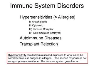
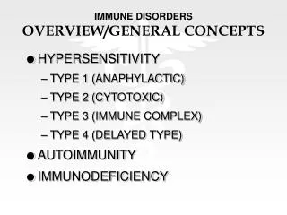
Hypersensitivities • Hypersensitivity • Any immune response against a foreign antigen exaggerated beyond the norm • Four types • Type I (immediate) • Type II (cytotoxic) • Type III (immune complex–mediated) • Type IV (delayed or cell-mediated)
Hypersensitivities • Type I (Immediate) Hypersensitivity • Localized or systemic reaction that results from the release of inflammatory molecules in response to an antigen • Develops within seconds or minutes following exposure to an antigen • Commonly called allergies • The antigens that stimulate it are called allergens
Figure 18.1a The mechanisms of a type I hypersensitivity reaction. Allergen (antigen) 1 Antigen-presenting cell (APC) phagocytizes and processes antigen. 2 APC presents epitope to Th2 cell. Th2 cell IL-4 3 IL-4 from Th2 cell stimulates selected B cell clone. B cell 4 B cells become plasma cells that secrete IgE. Plasma cell IgE against allergen 5 IgE stem binds to mast cells, basophils, and eosinophils. IgE Mast cell Eosinophil Basophil Sensitization
Figure 18.1b The mechanisms of a type I hypersensitivity reaction. Subsequent exposure to allergen Sensitized mast cell, basophil, or eosinophil Histamines, kinins, proteases, leukotrienes, prostaglandins, and other inflammatory molecules Degranulation
Hypersensitivities • Type I (Immediate) Hypersensitivity • The roles of degranulating cells in an allergic reaction • Mast cells • Distributed throughout the body in connective tissue • Have granules that contain inflammatory chemicals • Degranulation releases histamine, kinins, proteases, leukotrienes, and prostaglandins
Hypersensitivities • Type I (Immediate) Hypersensitivity • The roles of degranulating cells in an allergic reaction • Basophils • Least numerous leukocyte in the blood • Have granules that contain inflammatory chemicals • Degranulate like mast cells when encounter allergens
Hypersensitivities • Type I (Immediate) Hypersensitivity • The roles of degranulating cells in an allergic reaction • Eosinophils • Eosinophilia is the accumulation of eosinophils in blood • Mast cell degranulation can trigger the release of eosinophils from the bone marrow • Eosinophils in the bloodstream can degranulate • Release large amounts of leukotrienes • Increases severity of a hypersensitivity response
Hypersensitivities • Type I (Immediate) Hypersensitivity • Clinical signs of localized allergic reactions • Usually mild and localized • Site of reaction depends on portal of entry • Inhaled allergens may cause hay fever • Small inhaled allergens may reach lungs and cause asthma • Some allergens may cause inflammation of the skin, called hives or urticaria
Hypersensitivities • Type I (Immediate) Hypersensitivity • Clinical signs of systemic allergic reactions • Many mast cells may degranulate at once • Release large amounts of histamine and inflammatory mediators • Acute anaphylaxis or anaphylactic shock can result • Clinical signs are those of suffocation • Must be treated promptly with epinephrine • Common causes include bee stings and certain foods
Hypersensitivities • Type I (Immediate) Hypersensitivity • Diagnosis of type I hypersensitivity • Based on detection of high levels of allergen-specific IgE • Test referred to as ImmunoCAP specific IgE blood test, CAP RAST, or Pharmacia CAP • Can also diagnose using skin tests
Figure 18.4 Skin tests for diagnosing type I hypersensitivity.
Hypersensitivities • Type I (Immediate) Hypersensitivity • Prevention of type I hypersensitivity • Identification and avoidance of allergens • Food allergens are identified using an elimination diet • Immunotherapy ("allergy shots") can help prevent allergic reactions • Administration of a series of injections of dilute allergen • Must be repeated every two to three years • Not effective in treating asthma
Hypersensitivities • Type I (Immediate) Hypersensitivity • Treatment of type I hypersensitivity • Administer drugs that counteract inflammatory mediators • Antihistamines neutralize histamine • Treat asthma with a glucocorticoid and a bronchodilator • Epinephrine neutralizes many mechanisms of anaphylaxis • Relaxes smooth muscle • Reduces vascular permeability • Used in emergency treatment of severe asthma and anaphylactic shock
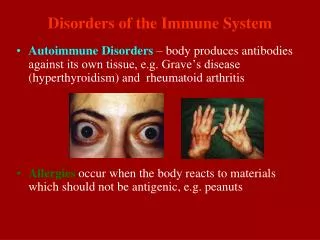
Hypersensitivities • Type II (Cytotoxic) Hypersensitivity • Results when cells are destroyed by an immune response • Often the combined activities of complement and antibodies • A component of many autoimmune diseases • Two significant examples • Destruction of blood cells following an incompatible blood transfusion • Destruction of fetal red blood cells
Hypersensitivities • Type II (Cytotoxic) Hypersensitivity • The ABO system and transfusion reactions • Blood group antigens are surface molecules of red blood cells • Each person's red blood cells have A antigen, B antigen, both antigens, or neither antigen • Transfusion reaction can result if an individual receives different blood type • Donor's blood group antigens may stimulate the production of antibodies in the recipient that destroy the transfused cells
Hypersensitivities • Type II (Cytotoxic) Hypersensitivity • The ABO system and transfusion reactions • Recipient has preexisting antibodies to foreign blood group antigens • Immediate destruction of donated blood cells can occur • Recipient has no preexisting antibodies to foreign blood group antigens • Transfused cells initially circulate and function normally • Eventually recipient's immune system mounts a primary response against the foreign antigens and destroys them
Figure 18.5 Events leading to hemolysis. Type A antigens on red blood cells of patient Donated red blood cells with B antigen Anti-B antibody Transfusion 1 Complement Hemoglobin Agglutination and complement binding 2 3 Hemolysis
Hypersensitivities • Type II (Cytotoxic) Hypersensitivity • The Rh system and hemolytic disease of the newborn • Rh antigen • Common to red blood cells of humans and rhesus monkeys • About 85% of humans are Rh-positive (Rh+) • If Rh−woman is carrying an Rh+ fetus, the fetus may be at risk for hemolytic disease • Administration of anti-Rh immunoglobulin, called RhoGAM, has reduced cases of hemolytic disease of the newborn
Figure 18.6 Events in the development of hemolytic disease of the newborn. During delivery, Rh antigens enter mother's circulation throughbreaks in the placenta. Mother makes anti-Rh antibodies, which have no effect on thisbaby, who has been born. Rh-negative mother Anti-Rh antibodies Placenta Rh+ antigens Fetal RBC with Rh+ antigens Rh-positive fetus Mother's Rh– RBC First pregnancy Mother's anti-Rh antibodies cross the placenta and destroy fetal blood cells. Mother's anti-Rh antibodies still circulate. Anti-Rh antibodies Placenta Fetal RBC with Rh+ antigens Fetal red blood cells destroyed Rh-positive fetus Subsequent pregnancy
Hypersensitivities • Type II (Cytotoxic) Hypersensitivity • Drug-induced cytotoxic reactions • Some drugs are too small to trigger immune responses • May become antigenic if bind to larger molecules • Can produce various diseases • Immune thrombocytopenic purpura • Agranulocytosis • Hemolytic anemia
Figure 18.7 Events in the development of immune thrombocytopenic purpura. Drug Platelet Drug molecules bind to platelets, forming drug-platelet complex. 1 Drug-platelet complex Drug-platelet complexes are antigenic, triggering an antibody immune response. 2 Antibody Antibodies bind to drug molecules; complement binds to antibodies. 3 Complement Membrane attacks complexes of complement lyse platelet, which leaks cytoplasm. 4
Hypersensitivities • Type III (Immune Complex–Mediated) Hypersensitivity • Caused by formation of immune complexes • Triggers release of inflammatory chemicals • Can cause localized reactions • Hypersensitivity pneumonitis • Glomerulonephritis • Can cause systemic reactions • Systemic lupus erythematosus • Rheumatoid arthritis
Figure 18.8 The mechanism of type III (immune complex-mediated) hypersensitivity. 1 Antigens combine with antibodies to form antigen-antibody complexes. Antigen-antibody complexes and activated complement attract and activate neutrophils, which release inflammatory chemicals. 4 Antigen Antibody (IgG) Neutrophil Antigen-antibody complex Inflammatory chemicals 2 Phagocytes remove most of the complexes, but some lodge in the walls of blood vessels. 5 Inflammatory chemicals damage underlying blood vessel wall. There the complexes activate complement. 3 Inactive complement Active complement
Hypersensitivities • Type III (Immune Complex–Mediated) Hypersensitivity • Hypersensitivity pneumonitis • Inhalation of antigens deep in the lungs stimulates the production of antibodies • Subsequent inhalation of the same antigen stimulates the formation of immune complexes • Activates complement
Hypersensitivities • Type III (Immune Complex–Mediated) Hypersensitivity • Glomerulonephritis • Immune complexes circulating in the bloodstream are deposited in the walls of glomeruli (small blood vessels in the kidneys) • Damage to the glomerular cells impedes blood filtration • Kidney failure and ultimately death result
Hypersensitivities • Type III (Immune Complex–Mediated) Hypersensitivity • Rheumatoid arthritis (RA) • Immune complexes are deposited in the joint • Results in release of inflammatory chemicals • The joints begin to break down and become distorted • Damage is progressively more severe • Trigger is not well understood • Treated with anti-inflammatory drugs
Figure 18.9 The crippling distortion of joints characteristic of rheumatoid arthritis.
Autoimmune Diseases • Type III (Immune Complex–Mediated) Hypersensitivity • Systemic lupus erythematosus (SLE) • Autoantibodies against DNA cause immune complex formation • Can cause glomerulonephritis and kidney failure • Many other autoantibodies can also occur • Against red blood cells, platelets, lymphocytes, muscle cells • Trigger unknown • Treatment with immunosuppressive drugs reduces autoantibody formation • Treatment with corticosteroids reduces inflammation
Figure 18.10 The characteristic facial rash of systemic lupus erythematosus.
Hypersensitivities • Type IV (Delayed or Cell-Mediated) Hypersensitivity • Inflammation occurs 12 to 24 hours after contact with certain antigens • Results from the actions of antigen, antigen-presenting cells, and T cells • Delay reflects the time it takes for macrophages andT cells to migrate to and proliferate at the site of the antigen
Hypersensitivities • Type IV (Delayed or Cell-Mediated) Hypersensitivity • The tuberculin response • Skin exposed to tuberculosis or tuberculosis vaccine reacts to an injection of tuberculin beneath the skin • Used to diagnose contact with antigens of M. tuberculosis • No response when individual has not been infected or vaccinated • Red, hard swelling develops in individuals previously infected or immunized • Response mediated by memory T cells, causing a slowly developing inflammation
Figure 18.11 A positive tuberculin test, a type IV hypersensitivity response.
Hypersensitivities • Type IV (Delayed or Cell-Mediated) Hypersensitivity • Allergic contact dermatitis • Cell-mediated immune response resulting in an intensely irritating skin rash • Triggered by chemically modified skin proteins that the body regards as foreign • In severe cases, acellular, fluid-filled blisters develop • Can be caused by poison ivy, formaldehyde, cosmetics, and chemicals used to produce latex • Treated with corticosteroids
Figure 18.12 Allergic contact dermatitis, a type IV hypersensitivity response.
Hypersensitivities • Type IV (Delayed or Cell-Mediated) Hypersensitivity • Graft rejection • Rejection of tissues or organs that have been transplanted • Grafts perceived as foreign by a recipient undergo rejection • Normal immune response against foreign MHC proteins present on graft cells • Likelihood of rejection depends on the degree to which the graft is foreign to the recipient • Based on the type of graft
Hypersensitivities • Type IV (Delayed or Cell-Mediated) Hypersensitivity • Graft-versus-host disease • Donated bone marrow cells regard the patient's cells as foreign • Donor and recipient differ in MHC class I molecules • Grafted T cells attack all of the recipient's tissues • Donor and recipient differ in MHC class II molecules • Grafted T cells attack the host's antigen-presenting cells • Immunosuppressive drugs can stop graft-versus-host disease
Hypersensitivities • Type IV (Delayed or Cell-Mediated) Hypersensitivity • Donor-recipient matching and tissue typing • MHC compatibility between donor and recipient is difficult because of high degree of variability • The more closely the donor and recipient are related, the smaller the difference in their MHC • Preferable that grafts be donated by a parent or sibling • Tissue typing is used to match donor and recipient
Hypersensitivities • Type IV (Delayed or Cell-Mediated) Hypersensitivity • The actions of immunosuppressive drugs • Immunosuppressive drugs are important to success of modern transplantation • Important classes of immunosuppressive drugs • Glucocorticoids • Cytotoxic drugs • Cyclosporine • Lymphocyte-depleting therapies
Hypersensitivities • Tell Me Why • During the war in Afghanistan in 2012, an army corporal with type AB blood received a life-saving blood transfusion from his sergeant, who had type O blood. Later the sergeant was involved in a traumatic accident and needed blood desperately. The corporal wanted to help but was told his blood was incompatible. Explain why the corporal could receive blood but could not give blood to the sergeant.
Autoimmune Diseases • Causes of Autoimmune Diseases • Occur more often in the elderly • Are more common in women than men • May result when an individual begins to make antibodies or cytotoxic T cells against normal body cells
Autoimmune Diseases • Causes of Autoimmune Diseases • Estrogen may stimulate destruction of tissue by cytotoxic T cells • Some maternal cells may cross the placenta, colonize the fetus, and trigger autoimmune disease later in life • Fetal cells may cross the placenta and trigger autoimmunity in the mother • Environmental factors such as viral infections • Genetic factors such as certain MHC genes • T cells may encounter self-antigens that are normally "hidden" • Microorganisms may trigger autoimmunity due to molecular mimicry • Failure of the normal control mechanisms of the immune system
Autoimmune Diseases • Examples of Autoimmune Diseases • Two major categories • Systemic autoimmune diseases • Single-organ autoimmune diseases • Can affect many different organs • Blood cells • Endocrine organs • Nervous tissue • Connective tissue