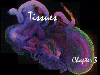
Understanding Tissues and Intercellular Connections in the Human Body
This chapter explores the fundamental tissue types in the human body and the mechanisms by which cells connect to form tissues. It details the three main intercellular connections: tight junctions, desmosomes, and gap junctions, and their roles in cell permeability and communication. The chapter classifies epithelial tissue based on its structure and function, including simple and stratified layers. Additionally, it covers connective tissues, muscle tissues, and their unique properties, emphasizing functions such as protection, support, and movement, forming the basis for understanding human anatomy.

Understanding Tissues and Intercellular Connections in the Human Body
E N D
Presentation Transcript
Intercellular Connections • Individual cells connect to form tissues 3 ways: • Tight junctions- • Desmosome- adhesion between cells in spots. Allows from some permeability. • Gap junctions- cytoplasms of adjacent cells are connected through transport proteins. • Ions can pass freely through cells.
Tissue Types A tissue is a group of cells with a common structure & function The human body is composed of four main tissue types: • 1. • 2. Connective • 3. • 4. Nerve
Epithelial Tissue Epithelial Tissue
Characteristics • Always has a free (apical) surface exposed to outside or open space. • Has a basement membrane to anchor underlying tissue
Functions • Covers body surfaces • Protects • Absorbs • Excretes
Classified by Shape • Squamous – • Cuboidal – • Columnar –
Classified by Shape May occur in layers: • Simple – • Stratified – 2 or more layers • Pseudostratified– • Example – simple cuboidal • Example – stratified columnar
s Simple Squamous- Thin, flattened cells. Allow for diffusion and filtration. Line air sacs of lungs and walls of capillaries.
Simple cuboidal-single layer of cube shaped cells. Lines follicles of thyroid gland, kidneys and ducts of certain glands.
Simple columnar- single layer of elongated cells. Can contain cilia, used for protection and absorption in digestive tract.
Stratified squamous-Layers of squamous cells. Make up epidermis and line cavities exposed to external environment.
Stratified columnar- Several layers of columnar cells overlying cuboidal cells near the basement membrane.
Pseudostratified ciliated columnar- Appear stratified but are not. Often contain cilia and goblet cells which secrete mucus.
Pseudostratified ciliated columnarw/goblet cells- Line Respiratory passages to trap unwanted particles
Transitional tissue- Changes in response to change in tension. Line urinary bladder and urethra.
Glandular Epithelium • Specialized to secrete substances • Those that secrete substances into ducts that open onto a surface are • Those that secrete into tissues or bloodare
Classifying Glands by Structure • Simple- • Compound- duct that does branch before secretory portion.
Classifying Glands by Type of Secretions 3 types: • Small portions of cells • in secretions • No loss of cytoplasm Ex. – mammary glands in secretions • Ex. – pancreas
Classifying by Secretions • Secretions w/entire cells filled w/secretory products; ex. – sebaceous (oil) glands
Functions • 1. connects • 2. • 3. protects • 4. • 5. fills spaces
Functions • 6. stores fat • 7. • 8. protects against infection • 9. • 10.helps repair damaged tissue
Characteristics • 1. Consists of cells in a matrix (intercellular material) • 2. Cells some distance apart • 3. • 4.
Types of Fibers: • collagenous – composed of collagen (protein); have great tensile strength; slightly elastic; compose bones, tendons & ligaments
Types of Fibers - continued • elastic – composed of elastin (protein); very elastic but weaker; compose vocal cords & air passages of lungs
Types of Fibers - continued • Reticular – composed of very fine collagenous fibers.
Types of Cells 1. Fixed cells – stay in one place & have stable numbers; 2 types: • fibroblasts – large & star-shaped; most prevalent
Types of Cells - continued • mast cells – may release heparin (for blood clotting) & histamines (promotes allergic reactions & inflammation); usually located near blood vessel walls
Types of Cells - continued 2. Wandering cells – • macrophages – (Purple cells – macrophages, Green cells – T-lymphocytes)
Areolar tissue-binds the skin to underlying organs and under epithelium to provide bloodflow.
Adipose tissue- connective tissue composed of fats, cushion joints and provide insulation
Regular dense connective- strong fibers bind body parts together. Found in ligaments and tendons.
Irregular dense connective- disorganized and strong. Found in the dermis
Hyaline cartilage- Most common, found on ends of bones, nose cavity and supporting rings of resp. system.
Fibrocartilage- tough tissue containing collagenous fibers. Shock absorbers between vertebrae.
Elastic cartilage- flexible cartilage make up ears and larynx
Bone- A- central canal (contains blood vessels) B- Canaliculi- minute tubes allow for movement between cells.
Muscle Tissue 3 types: • Skeletal- • Used for movement • Smooth- lacks striations found in skeletal, used for involuntary movements • Ex- move food through digestive tract • Cardiac-