Download
1 / 65
650 likes | 715 Views
Explore nucleotides, nucleosides, and nitrogen bases in DNA and RNA, understand cAMP and ADP structures, functions in biology and cellular energetics, as well as genetic codes.

E N D
Name: Dr. Pramod B. ThakurClass: M.Sc - II Subject: Organic Chemistry Title of Topic: Nucleic acids
Msc II -Organic Chemistry Course code – PSCHO304, paper-4, Unit-22.2 Nucleic acids (Total 06 Lectures) 1. Structure and Function of Nucleotide- c-AmP 2. Structure and Function of Nucleotide- ADP 3. Structure and Function of Nucleotide- ATP 4. Structure and Function of Nucleic acid- DNA 5. Structure and Function of Nucleic acid- RNA 6. Replication 7. Genetic code
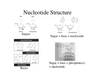
Nucleotide • A molecule consisting of a heterocyclic base, a pentose sugar unitand a phosphate group is called nucleotide. • Aheterocyclic base can have purine (Adenine, Guanine) or pyrimidine(Uracil, Cytosine, Thymine) structure. • A pentose sugar unit can have ribose or deoxyribose. • phosphate group contain phosporic acid molecular unit. • The sugaris linked to the base via an -N-glycosidic bond. • Nucleotides are perhaps best known as the monomericunits of DNA and RNA. • A molecule consisting of only the base and the sugar unit is called a nucleoside. • Hence nucleotide are also known as phosphate ester of nucleoside .
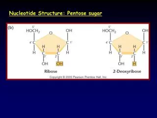
DEOXYRIBOSE RIBOSE CH2OH CH2OH OH OH O O C C C C H H H H H H H H C C C C OH OH H OH A pentose sugar unit
Building Blocks * Nucleotides = Base + Sugar + Phosphate * Nucleosides = Base + Sugar * Nitrogen Bases • Purines (5 + 6 membered rings) • Adenine • Guanine • Pyrimidines (6 membered ring) • Thymine • Cytosine • Uracil *Pentose Sugars (numbering) 1. Ribose 2. Deoxy Ribose * Phosphoric acis
Nucleotides occur not only as mono phosphates but also as di and triphosphates.
AMP, ADP and ATP • Additional phosphate groups can be added to the nucleoside 5’-monophosphates to form diphosphates and triphosphates
NH2 N N N N CH2 O O O OH P O HO 1.Structure and Function of Nucleotide- c-AmP Structure of c-AmP: Cyclic adenosine monophosphate (cAMP, cyclic AMP or 3'-5'-cyclic adenosine monophosphate) Cyclic adenosine monophosphate (cAMP)
c-AMP synthesis • cAMP is synthesised from ATP by adenylatecyclase located on the inner side of the plasma membrane. • Adenylatecyclase is activated by a range of signaling molecules through the activation of adenylatecyclase stimulatory G (Gs)-protein-coupled receptors and inhibited by agonists of adenylatecyclase inhibitory G (Gi)-protein-coupled receptors. • Liver adenylatecyclase responds more strongly to glucagon, and muscle adenylatecyclase responds more strongly to adrenaline.
Important biological functions of - c-AmP: • cAMP is a second messenger, used for intracellular signal transduction, such as transferring into cells the effects of hormones like glucagon and adrenaline, which cannot pass through the cell membrane. • It is involved in the activation of protein kinases and regulates the effects of adrenaline and glucagon. • cAMP also binds to and regulates the function of ion channels such as the HCN channels and a few other cyclic nucleotide-binding proteins such as Epac1 and RAPGEF2. • cAMP and its associated kinases function in several biochemical processes, including the regulation of glycogen, sugar and lipid metabolism. In eukaryotes, cyclic AMP works by activating protein kinase A. • In the species Dictyostelium discoideum, cAMP acts outside the cell as a secreted signal.
Some research has suggested that a deregulation of cAMP pathways and an aberrant activation of cAMP controlled genes is linked to the growth of some cancers. • cAMP receptor protein increases expression of a large number of genes, including some encoding enzymes that can supply energy independent of glucose.
NH2 N N O O N N OCH2 HO O P P O HO HO HO OH 2.Structure and Function of Nucleotide- ADP Structure of ADP: Adenosine Diphosphate (ADP)
Structure of Adenosine Diphosphate (ADP): • Heterocyclic Base Unit : adenine • Pentose Sugar Unit : Ribose • Phosphate Unit : 2 Phosphate unit • A molecule of ADP consists of three important structural components: a sugar backbone which is attached to a molecule of adenine and two phosphate groups bonded to the 5' carbon atom of ribose. • The two phosphate groups of ADP are added in series to the 5’ carbon of the sugar backbone, while the adenosine molecule attaches to the 1’carbon.
Adenosine Diphosphate (ADP): Additional phosphate groups can be added to the nucleoside 5’-monophosphates to form diphosphates
Important biological functions of - ADP: • Bioenergetics: ADP-ATP cycling supplies the energy needed to do work in a biological system. • Eg. It takes multiple reactions to effectively produce one muscle contraction and therefore the availability of large amounts of ATP is required to produce each muscle contraction. • Breaking one of ATP’s phosphorus bonds generates approximately 31 kilojoules per Mole of ATP. ADP can be converted, or powered back to ATP through the process of releasing the chemical energy available in food.
2. Cellular respiration: * Catabolism- ADP and phosphate are needed as precursors to synthesize ATP. The enzymes phosphoglycerate kinase and pyruvate kinase facilitate the addition of a phosphate group to ADP. * Glycolysis- Glycolysis is performed by all living organisms which uses energy obtained from the hydrolysis of ATP to ADP. *Citric acid cycle: actively involved in Citric acid cycle (GTP + ADP → GDP + ATP) * Oxidative phosphorylation: Oxidative phosphorylation produces 26 of the 30 molecules of ATP generated in cellular respiration by transferring electrons from NADH or FADH2 to O2 through electron carriers.The energy released when electrons are passed from higher energy NADH or FADH2 to the lower energy O2 is required to phosphorylate ADP and once again generate ATP. It is this energy coupling and phosphorylation of ADP to ATP, that gives the electron transport chain the name oxidative phosphorylation.
3. Mitochondrial ATP synthase complex: • Actively involved in The ATP synthase complex exists both within the mitochondrial membrane and protrudes into the matrix. • The energy derived as a result of the chemical gradient is then used to synthesize ATP by coupling the reaction of inorganic phosphate to ADP in the active site of the ATP synthase enzyme; • The equation for this can be written as ADP +Pi → ATP. • 4. Blood platelet activation: • Normally, small disk shaped platelets circulate the blood freely and without interaction with one another. • ADP is stored in dense bodies inside blood platelets and is released upon platelet activation. • ADP interacts with a family of ADP receptors found on platelets which leads to platelet activation.
NH2 N N O O O N N HO OCH2 P O O P P O HO HO HO HO OH 3.Structure and Function of Nucleotide- ATP Structure of ATP: Adenosine-5'-triphosphate (ATP)
Structure of Adenosine-5'-triphosphate (ATP): • Heterocyclic Base Unit : adenine • Pentose Sugar Unit : Ribose • Phosphate Unit : 3 Phosphate unit • A molecule of ADP consists of three important structural components: a sugar backbone which is attached to a molecule of adenine and Three phosphate groups bonded to the 5' carbon atom of ribose. • The three phosphate groups of ATP are added in series to the 5’ carbon of the sugar backbone, while the adenosine molecule attaches to the 1’carbon. • The phosphoryl groups, starting with the group closest to the ribose, are referred to as the alpha (α), beta (β), and gamma (γ) phosphates. • ATP is highly soluble in water and is quite stable in solutions between pH 6.8–7.4, but is rapidly hydrolysed at extreme pH. Consequently, ATP is best stored as an anhydrous salt .
Structure of Adenosine-5'-triphosphate (ATP): • Additional phosphate groups can be added to the nucleoside 5’-monophosphates to form triphosphates
Important biological functions of - ATP: Metabolism: • ATP is the major energy source for cellular activity. • ATP is consumed in the cell by energy-requiring (endothermic) processes and can be generated by energy releasing (exothermic) processes. In this way ATP transfers energy between spatially separate metabolic reactions. hence It is often called the "molecular unit of currency" of intracellular energy transfer. Synthesis: • ATP is directly involved in the synthesis of macromolecules, including DNA and RNA and proteins. Active Transport: 1. ATP also plays a critical role in the transport of macromolecules across cell membranes, e.g. exocytosis and endocytosis.
cell structure and locomotion: • ATP is critically involved in maintaining cell structure. Eg. ATP is required for the shortening of actin and myosin filament cross bridges required for muscle contraction. This latter process is one of the main energy requirements of animals and is essential for locomotion and respiration. Cell signalling: • ATP is critical in signal transduction processes. It is also a signalling molecule. In humans, this signalling role is important in both the central and peripheral nervous system. 2. Kinase activity on substrates such as proteins or membrane lipids are a common form of signal transduction. 3. In the CNS, it has multiple functions, such as modulation of neural development, neuron and glial signalling and the control of innate and adaptive immune systems.
ATP is also used by adenylate cyclase and is transformed to the second messenger molecule cyclic AMP, which is involved in triggering calcium signals by the release of calcium from intracellular stores. This form of signal transduction is particularly important in brain function, although it is involved in the regulation of a multitude of other cellular processes. Amino acid activation in protein synthesis: • The energy required for the activation of amino acid in protein synthesis is supplied from energy reliesed in ATP hydrolysis to adenosin monophosphate (AMP).
1.Structure and Function of Nucleic acid-DNA & RNA • Nucleic acid: • Friedrich Miescher in 1869 isolated what he called nucleinfrom the nuclei of pus cells Nuclein was shown to have acidic properties, hence it became called nucleic acid. • Two types of nucleic acid are found 1. Deoxyribonucleic acid (DNA) 2. Ribonucleic acid (RNA) • DNA is found in the nucleus in the eukaryotic cell with small amounts in mitochondria and chloroplasts. • RNA is found throughout the cell. • Nucleic acids are polynucleotides. • Their building blocks are nucleotides.
Primary Structure of Nucleic Acids • The primary structure of a nucleic acid is the nucleotide sequence • The nucleotides in nucleic acids are joined by phosphodiester bonds • The 3’-OH group of the sugar in one nucleotide forms an ester bond to the phosphate group on the 5’-carbon of the sugar of the next nucleotide
Reading Primary Structure • A nucleic acid polymer has a free 5’-phosphate group at one end and a free 3’-OH group at the other end • The sequence is read from the free 5’-end using the letters of the bases • This example reads 5’—A—C—G—T—3’
Example of DNA Primary Structure • In DNA, A, C, G, and T are linked by 3’-5’ ester bonds between deoxyribose and phosphate
Secondary Structure Of DNA Hydrogen bonds T T A C C G P P P C P P G P P G P P T P A P A P Structural Basis of Chargaff’s Rules • Two Strands have complementary sequences • 2 logical operations to obtain complementary strand 5' to 3' • 1. Reverse: Rewrite the sequence, back to front • 2. Complement: Swap A with T, C with G James D. Watson and Francis H. C. Crick proposed a structure for DNA in 1953. Watson and Crick's structure was based on:•Chargaff's observations•X-ray crystallographic data of Maurice Wilkins and Rosalind Franklin •Model building
4. Structure and Function of Nucleic acid- DNA Structure Of DNA: • DNA IS MADE OF TWO STRANDS OF POLYNUCLEOTIDE • The sister strands of the DNA molecule run in opposite directions (antiparallel- 3’-5’) • They are joined by the bases. • Each base is paired with a specific partner: Purine with Pyrimidine A is always paired with T G is always paired with C • This the sister strands are complementary but not identical • The bases are joined by hydrogen bonds, individually weak but collectively strong. • Bases are very nearly planar.
2-deoxyribose 2-deoxyribose A T Base Pairing • Watson and Crick proposed that A and T were present in equal amounts in DNA because of complementary hydrogen bonding.
2-deoxyribose 2-deoxyribose G C Base Pairing • Likewise, the amounts of G and C in DNA were equal because of complementary hydrogen bonding.
Two antiparallel strands of DNA are paired by hydrogen bonds between purine and pyrimidine bases.
Model of DNA: • The model was developed by Watson and Crick in 1953. • They received a nobel prize in 1962 for their work. • The model looks like a twisted ladder – double helix.
Watson Crick Died in 2004
The sides of the ladder are: • P = phosphate • S = sugar molecule • The steps of the ladder are C, G, T, A = nitrogenous bases • (Nitrogenous means containing the element nitrogen.) • A = Adenine • T = Thymine • A always pairs with T in DNA • C = Cytosine • G = Guanine • C always pairs with G in DNA Nucleotide Untwisted it looks like this: (Apples are Tasty) (Cookies are Good)
the minor groove, is 12 Å wide the major groove, is 22 Å wide
Biological Function of Nucleic acid- DNA Genes and genomes: The genetic information in a genome is held within genes, and the complete set of this information in an organism is called its genotype. A gene is a unit of heredity and is a region of DNA that influences a particular characteristic in an organism. Transcription : In transcription, the codons of a gene are copied into messenger RNA by RNA polymerase. This RNA copy is then decoded by a ribosome that reads the RNA sequence by base-pairing the messenger RNA to transfer RNA, which carries amino acids. Since there are 4 bases in 3-letter combinations, there are 64 possible codons( combinations). These encode the twenty standard amino acids, giving most amino acids more than one possible codon. There are also three 'stop' or 'nonsense' codons signifying the end of the coding region; these are the TAA, TGA and TAG codons.