Download
1 / 23
240 likes | 559 Views
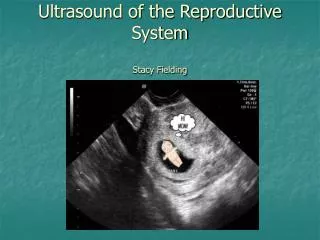
Ultrasound of the Reproductive System Stacy Fielding. Technique. 7.5-10mHz transducers 5 mHz in mid to late term pregnancy, pyometra, ovarian tumors Dorsal recumbency is routine Larger animals standing Full bladder enhances visualization of uterus Acoustic window. Ovaries.

E N D
Technique • 7.5-10mHz transducers • 5 mHz in mid to late term pregnancy, pyometra, ovarian tumors • Dorsal recumbency is routine • Larger animals standing • Full bladder enhances visualization of uterus • Acoustic window
Ovaries • Near caudal pole of kidney • 1.5cm x 0.7cm x 0.5cm • Homogenous parenchyma • Cortex and medulla • Hard to differentiate • Cortex contains follicles
Ovaries-cont’d • Anestrus/early proestrus • Small, oval to bean shaped • Homogenous echogenicity (like renal cortex) • Follicles anechoic, become larger as ovulation approaches • Diestrus • CL/CH has multifocal anechoic to hyperechoic areas
Cystic Ovaries Anechoic Thin walls Acoustic enhancement Solitary/multiple Unilateral/bilateral May see associated pyometra or hydrometra Ovarian Neoplasia Several kinds Mostly unilateral Smooth or irregular margins Variable appearance MET CHECK! Ovaries
Normal Uterus • Location: between urinary bladder and descending colon • Various sizes • Uterine horns: 10-14cm x 0.5-1.0cm • Uterine body: 1.4-3cm x 3cm • Cervix: 1.5-2cm x 0.8cm • Solid, homogenous, relatively hypoechoic • May have thin hyperechoic border • Cannot differentiate layers
Uterine horns not easily identified Lost in mesenteric fat and small bowel echoes Cervix Hyperechoic, linear Normal Uterus-cont’d
U/S is modality of choice for Dx Enlarged uterus & uterine horns Luminal contents Homogenous, anechoic Echogenic, “swirling” May see varying wall thickness Endometrium may contain anechoic foci Ddx: hydrometra, mucometra Monitor response to therapy Pyometra
Uterine Neoplasia • Adenomas • Leiomyomas • Leiomyosarcomas • Isoechoic to surrounding tissue, may project into the lumen
Pregnancy Diagnosis • 21-35 days after breeding • Remember conception can occur several days afterward • May be difficult to count fetuses • Radiography recommended late in gestation, following mineralization • Earliest change = enlarging uterus • Non-specific • Gestational sac (blastocyst) • First confirmatory sign • Anechoic, several mm diameter • Thin, hyperechoic wall • 17-20 d
Pregnancy-cont’d • Embryo: 23-25 d • Oblong, echogenic, several mm • Yolk sac: 25-28 d • U-shaped echogenic structure • Extends across gestational sac • Zonary placenta: 27-30d • Thin hyperechoic layer surrounding gestational sac
Cardiac activity Day 23-25 post LH surge Fluttering echoes Fetal movement Day 35 Fetal resorption before day 25 Embryonic fluid becomes hypoechoic Echogenic particles Abortion After day 35 Sonographic appearance disappears Fetal Viability
Caudal to bladder, at level of trigone Ventral to rectum Normal appearance varies Age Neuter status Young to middle age intact male: homogenous, moderate echogenicity Smooth margins, hyperechoic rim Prostatic urethra Hypoechoic to anechoic round structure on midline Prostate
Benign Prostatic Hyperplasia Older intact dogs Symmetrical enlargement May be up to 4 times normal Variable echogenicity and texture Prostatitis Acute or chronic Symmetrical or asymmetrical Heterogenous appearance May see hypoechoic areas (cyst or abscess) Mineralization, fibrosis Prostate Diseases
Neoplasia Older neutered dogs Enlarged Irregular shape Texture varies Hyperechoic foci with acoustic shadowing = mineralization May see cyst-like lesions Examine surrounding structures! Cysts Developmental or congenital Anechoic contents Thin hyperechoic wall Vary in size and # Paraprostatic Cysts Muellerian duct remnant or extension of lobe Anechoic, fluid filled May see swirlies Variable wall thickness May see compartments Prostate Diseases
Prostatic Cyst www.merckvetmanual.com
Testes • Homogenous texture • Parietal and visceral tunics: • hyperechoic • Mediastinum testis: • Echogenic linear structure on midline • Tail of the epididymis • Nearly anechoic • Coarse echotexture
Testicular Neoplasia • Interstitial, Sertoli cell, seminoma • May all appear the same • Mixed appearance on U/S • Hemorrhage • Necrosis • May obliterate mediastinum testis +/- epididymis
Retained testes Small size Caudal to kidneys to inguinal canal Difficult to see on U/S Orchitis Patchy, hypoechoic parenchyma Hyperechoic if chronic Epididymal enlargement Abscesses Irregular shaped Hypoechoic contents May look like neoplasia! Testes-cont’d