Chapter 12
Muscle: Mechanisms of Contraction and Neural Control. Chapter 12. Objectives. Describe the nature of a muscle twitch and explain how summation and tetanus are produced. Explain what is meant by the sliding filament theory of contraction.

Chapter 12
E N D
Presentation Transcript
Muscle: Mechanisms of Contraction and Neural Control Chapter 12
Objectives Describe the nature of a muscle twitch and explain how summation and tetanus are produced. Explain what is meant by the sliding filament theory of contraction. List the events that occur during cross-bridge cycles and describe the role of ATP in muscle contraction. Explain how tropomyosin and troponin control muscle contraction and relaxation, and describe the role of Ca2+ and the SR in excitation-contraction coupling.
Objectives (continued) Describe the structure and functions of muscle spindles and explain the mechanisms involved in a stretch reflex. Describe the functions of the Golgi tendon organs and explain why a slow, gradual muscle stretch could avoid the spasm that may result from a rapid stretch. Explain how slow-twitch, fast-twitch, and intermediate fibers differ in structure and function. Describe skeletal muscle metabolism during exercise, and explain how muscle fatigue and how muscle fibers change as a result of physical training.
Motor Unit When somatic neuron is activated, all the muscle fibers it innervates contract with all or none contractions. Innervation ratio: Ratio of motor neuron: muscle fibers. Fine neural control over the strength occurs when many small motor units are involved. Recruitment: Larger and larger motor units are activated to produce greater strength.
Motor Unit (continued) • Each somatic neuron together with all the muscle fibers it innervates. • Each muscle fiber receives a single axon terminal from a somatic neuron. • Each axon can have collateral branches to innervate an equal # of fibers. Figure 12-4
Mechanisms of Contraction • Thick filaments: • Primarily composed of myosin. • Thin filaments: • Primarily composed of actin. • Sarcomere: • Z disc to Z disc. • Titin: • Elastic protein that runs through the myosin from M line to Z disc. • Contributes to elastic recoil of muscle. Figure 12-8
Excitation-Contraction Coupling • Na+ diffusion produces end-plate potential (depolarization). • + ions are attracted to negative plasma membrane. • If depolarization sufficient, threshold occurs, producing APs. Figure 12-16
Excitation-Contraction Coupling (continued) • APs travel down sarcolema and T tubules. • SR terminal cisternae releases Ca2+ from chemical release channels: • Ca2+ is also released through a Ca2+-induced Ca2+ release. Figure 12-15
Excitation-Contraction Coupling (continued) • Ca2+ attaches to troponin. • Tropomyosin-troponin complex configuration change occurs. • Cross bridges attach to actin. • Each myosin head contains an ATP-binding site. • The myosin head functions as a myosin ATPase. • Myosin binding site splits ATP to ADP and Pi. • ADP and Pi remain bound to myosin until myosin heads attach to actin. Figure 12-10
Contraction • Pi is released, causing the power stroke to occur. • Power stroke pulls actin toward the center of the A band. • ADP is released, when myosin binds to a fresh ATP at the end of the power stroke. • Release of ADP upon binding to another ATP, causes the cross bridge bond to break. • Cross bridges detach, ready to bind again. • Synchronous action: • Only 50% of the cross bridges are attached at any given time. Figure 12-11
Contraction (continued) Figure 12-12
Sliding Filament Theory of Contraction Muscle contracts: Occurs because of sliding of thin filaments over and between thick filaments towards center. Shortening the distance from Z disc to Z disc. A bands: Contain actin. Move closer together. Do not shorten. I bands: Distance between A bands of successive sarcomeres. Decrease in length. H bands shorten. Contain only myosin. Shorten during contraction.
Regulation of Contraction Regulation of cross bridge attachment to actin due to: Tropomyosin:. Lies within grove between double row of G-actin. Troponin: Attached to tropomyosin. Serves as a switch for muscle contraction and relaxation. In relaxed muscle: Tropomyosin blocks binding sites on actin.
Muscle Relaxation Muscle Relaxation: [Ca2+] in sarcoplasm low when tropomyosin blocks attachment. Prevents muscle contraction. Ca2+ is pumped back into the SR in the terminal cisternae. Muscle relaxes. APs must cease for the muscle to relax. ACh-esterase degrades ACh. Ca2+ release channels close. Ca2+ pumped back into SR through Ca2+-ATPase pumps. Choline recycled to make more ACh.
Twitch, Summation, and Tetanus Twitch: Muscle is stimulated with a single electrical shock (above threshold). Quickly contracts and then relaxes. Increasing stimulus increases the strength of the twitch (up to maximum). Summation: If second electrical shock is administered before complete relaxation of muscle. Incomplete tetanus: Stimulator delivers an increasing frequency of electrical shocks. Relaxation period shortens between twitches. Strength of contraction increases.
Twitch, Summation, and Tetanus (continued) • Complete tetanus: • Fusion frequency of stimulation. • No visible relaxation between twitches. • Smooth sustained contraction. • Treppe: • Staircase effect. • Electrical shocks are delivered at maximum voltage. • Each shock produces a separate, stronger twitch (up to maximum). • Due to increase in intracellular Ca2+. • Represents “warm-up.”
Twitch, Summation, and Tetanus (continued) Figure 12-18
Isotonic, Isometric, and Eccentric Contractions In order for a muscle fiber to shorten, they must generate a force greater than the opposing forces that act to prevent movement of that muscle insertion. Isotonic contractions: Force of contraction remains constant throughout the shortening process. Velocity of muscle shortening decreases as load increases. Isometric contractions: Length of muscle fibers remain constant, if the number of muscle fibers activated is too few to shorten the muscle. Velocity of shortening is 0.
Isotonic, Isometric, and Eccentric Contractions (continued) • Force-velocity curve: • Inverse relationship between force opposing muscle contraction and velocity of muscle shortening. • Eccentric contractions: • Force exerted on a muscle to stretch, it is greater than the force of muscle contraction. • Muscle will lengthen as it contracts. Figure 12-19
Series-Elastic Component Non-contractile tendons and connective tissue absorb tension as the muscle contracts. Tendons first must be pulled tight, before the muscle contraction results in shortening. Tendons: Have elasticity (resist distension). Display recoil. Spring back to resting length.
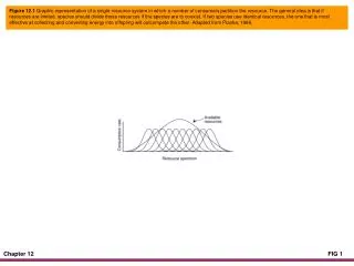
Length-Tension Relationship • Strength of muscle contraction influenced by: • Frequency of stimulation. • Thickness of each muscle fiber. • Initial length of muscle fiber. • Ideal resting length: • Length which can generate maximum force. • Overlap too small: • Few cross bridges can attach. • No overlap: • No cross bridges can attach to actin. Figure 12-20
Neural Control of Skeletal Muscles Lower motor neuron activity influenced by: Sensory feedback from the muscles and tendons. Facilitory and inhibitory effects of upper motor neurons. Cell bodies in spinal cord and axons within neurons that stimulate muscle contractions. Final common pathway by which sensory stimuli and higher brain centers exert control over skeletal movements.
Muscle Spindle Apparatus To control skeletal muscle movements, it must receive continuous sensory feedback. Sensory feedback includes information from: Golgi tendon organs: Sense tension that the muscle exerts on the tendons. Muscle spindle apparatus: Measures muscle length. Contains thin muscle cells called intrafusal fibers. Contractile apparatus absent from central regions. 2 types of intrafusal fibers: Nuclear bag fibers: Nuclei arranged in loose aggregate. Nuclear chain fibers: Nuclei arranged in rows.
Muscle Spindle Apparatus (continued) Sensory neurons: Primary, annulospiral sensory endings: Wrap around the central regions of both nuclear bag and chain fibers. Most stimulated at onset of stretch. Secondary, flower-spray endings: Located over the contracting poles of nuclear chain fibers. Respond to tonic (sustained) stretch. Sudden, rapid stretching of a muscle causes spindles to stretch, stimulating both primary and secondary endings Produces more forceful muscle contraction.
Muscle Spindle Apparatus (continued) Extrafusal fibers: Ordinary muscle fibers outside the spindles. Contain myofibrils along entire length. Spindles are arranged in parallel with the extrafusal muscle fibers. Only extrafusal muscle fibers are strong and numerous enough to cause muscle contraction.
Alpha and Gamma Motor Neurons • 2 types of lower motor neurons in the spinal cord: • motor neurons: • Neurons that innervate extrafusal fibers. • Fast conducting fibers. • motor neurons: • Neurons that innervate the intrafusal fibers. • Cause isometric muscle contraction. • Too few in # to cause muscle to shorten. • Stimulation by a motor neurons only, can cause skeletal muscle movements. Figure 12-26
Coactivation of Alpha and Gamma Motor Neurons Coactivation: Upper motor neurons usually stimulate and motor neurons simultaneously. Stimulation of motor neurons results in muscle contraction and shortening. Stimulation of motor neurons stimulate intrafusal fibers and take out the slack. Activity of motor neurons is maintained to keep muscle spindles under proper tension while muscles are relaxed.
Monosynaptic-Stretch Reflex • Consists of only one synapse within the CNS. • Sensory neuron synapses directly with the motor neuron. • Striking the patellar ligament, passively stretches the spindles. • Stimulates primary endings in spindles, activating sensory neurons. • Synapse withmotor neurons stimulating extrafusal fibers. • Produces isotonic contraction, (knee jerk). Figure 12-27
Golgi Tendon Organ Reflex • Disynaptic reflex. • 2 synapses are crossed in the CNS. • Sensory neurons synapse with interneurons. • These interneurons have inhibitory synapses with motor neurons. • Helps prevent excessive muscle contraction or passive muscle stretching. Figure 12-28
Reciprocal Innervation • Sensory neuron stimulates motor neuron and interneuron. • Interneurons inhibit motor neurons of antagonistic muscles. • When limb is flexed, antagonistic extensor muscles are passively stretched. Figure 12-29
Crossed-Extensor Reflex • Double reciprocal innervation. • Affects muscles on the contralateral side of the cord. • Step on tack: • Foot is withdrawn by contraction of flexors and relaxation of extensors. • Contralateral leg extends to support body. Figure 12-30
Upper Motor Neuron Control of Skeletal Muscles Influence lower motor neurons. Pyramidal tracts: Neurons in precentral gyrus contribute axons that cross to contralateral sides in the pyramids of medulla. Extrapyramidal tracts: Neurons in the other areas of the brain.
Upper Motor Neuron Control of Skeletal Muscles (continued) Cerebellum: Receives sensory input from muscle spindles, Golgi tendon organs, and areas of cerebral cortex devoted to vision, hearing and equilibrium. No descending tracts from the cerebellum. Influences motor activity indirectly. All output from cerebellum is inhibitory. Aids motor coordination. Basal nuclei: Include caudate nucleus, putamen, globus pallidus, and nuclei of thalamus, substantia nigra and red nucleus. Profound inhibitory effects on the activity of lower motor neurons. Damage to basal nuclei result in increased muscle tone.
Slow- and Fast-Twitch Fibers Skeletal muscle fibers can be divided on basis of contraction speed: Slow-twitch (type I fibers). Fast-twitch (type II fibers). Differences due to different myosin ATPase isoenzymes that are slow or fast.
Slow- and Fast-Twitch Fibers (continued) Slow-twitch (type I fibers): Red fibers. High oxidative capacity for aerobic respiration. Resistant to fatigue. Have rich capillary supply. Numerous mitochondria and aerobic enzymes. High [myoglobin]. Soleus muscle in the leg.
Slow- and Fast-Twitch Fibers (continued) Fast-twitch (type IIX fibers): White fibers. Adapted to respire anaerobically. Have large stores of glycogen. Have few capillaries. Have few mitochondria. Extraocular muscles that position the eye. Intermediate (type II A) fibers: Great aerobic ability. Resistant to fatigue. People vary genetically in proportion of fast- and slow-twitch fibers in their muscles.
Muscle Fatigue Any exercise induced reduction in the ability to maintain muscle to generate force or power. Sustained muscle contraction fatigue is due to an accumulation of ECF K+. Repolarization phase of AP. During moderate exercise fatigue occurs when slow-twitch fibers deplete their glycogen reserve. Fast twitch fibers are recruited, converting glucose to lactic acid. Interferes with Ca2+ transport. Central fatigue: Muscle fatigue caused by changes in CNS rather than fatigue of muscles themselves.
Muscle Fuel Consumption During Exercise Figure 12-21
Metabolism of Skeletal Muscles Skeletal muscles respire anaerobically first 45 - 90 sec of moderate to heavy exercise. Cardiopulmonary system requires this amount of time to increase 02 supply to exercising muscles. If exercise is moderate, aerobic respiration contributes the majority of skeletal muscle requirements following the first 2 min. of exercise. Maximum oxygen uptake (aerobic capacity): Maximum rate of oxygen consumption (V02 max) determined by age, gender, and size.
Metabolism of Skeletal Muscles Lactate threshold: % of max. 02 uptake at which there is a significant rise in blood [lactate]. Healthy individual, significant blood [lactate] appears at 50– 70% V02 max. During light exercise: Most energy is derived from aerobic respiration of fatty acids. During moderate exercise: Energy is derived equally from fatty acids and glucose. During heavy exercise: Glucose supplies 2/3 of the energy for muscles. Liver increases glycogenolysis. During exercise, the GLUT-4 carrier protein is moved to the muscle cell’s plasma membrane.
Metabolism of Skeletal Muscles (continued) Oxygen debt: Oxygen that was withdrawn from hemoglobin and myoglobin during exercise. Extra 02 required for metabolism tissue warmed during exercise. 02 needed for metabolism of lactic acid produced during anaerobic respiration. When person stops exercising, rate of oxygen uptake does not immediately return to pre-exercise levels. Returns slowly.
Metabolism of Skeletal Muscles (continued) • Phosphocreatine (creatine phosphate): • Rapid source of renewal of ATP. • ADP combines with creatine phosphate. • [Phosphocreatine] is 3 times [ATP]. • Ready source of high-energy phosphate. Figure 12-22
Adaptations of Muscles to Exercise Training Maximum 02 uptake during strenuous exercise: In adult aged 20-25, averages 50 ml of 02/min. In trained endurance athlete increases up to 86 ml of 02/min. Increases lactate threshold. Produces less lactic acid. Increases proportion of energy derived from aerobic respiration of fatty acids. Lowers depletion of glycogen stores.
Adaptations of Muscles to Exercise Training (continued) All fibers adapt to endurance training: Increase # of mitochondria. Endurance training produces an increase in type IIA fibers and a decrease in type IIX fibers. Does not increase size of muscles. Muscle enlargement produced by: Frequent periods of high-intensity exercise in which muscles work against high-resistance. Type II fibers become thicker. May split into 2 myofibrils.