Download
1 / 42
470 likes | 1.54k Views
Skin Scrapes and Their Parasitic Friends. Clinical Pathology. Skin Scrape. Fast and easy diagnostic tool Inexpensive Should be one of the first diagnostics performed when diagnosing dermatologic disorders. Items needed for a skin scrape. Blade (15 or 10) Mineral oil Microscope slide

E N D
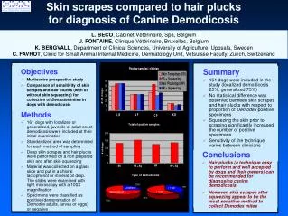
Skin Scrapes and Their Parasitic Friends Clinical Pathology
Skin Scrape • Fast and easy diagnostic tool • Inexpensive • Should be one of the first diagnostics performed when diagnosing dermatologic disorders.
Items needed for a skin scrape • Blade (15 or 10) • Mineral oil • Microscope slide • Microscope
Skin Scraping Technique • Drop mineral oil on skin and/or slide. • Some prefer just to pass blade through oil instead of applying to skin. • Gently squeeze the skin area that you are scraping. Hold the blade perpendicular to the skin. • Scrape until a small of amount of capillary blood oozes. (careful to not cut). • May place coverslip if you like. • Examine entire slide in a systematic fashion (start with 10x objective)
Skin Scrape Hints • Scrape areas that have lesions • Scrape typical sites for particular ectoparasite • Ex: ear margins for Sarcoptic mange • Do multiple skin scrapes
Microscopic exam of the Skin Scrape Sample • Identify any ectoparasites. • Determine live:dead ratios • Determine life cycle stage • Eggs, immature, adults • Determine numbers found
Classification • Class • Acarina (mites and ticks) • Family: Sarcoptidae and Psoroptidae • Sarcoptidae • Mites that burrow through epidermis • Sarcoptes, Notoedres, Knemidocoptes species • Psoroptidae • Mites that reside on the skin surface • Psoroptes, Chorioptes, Otodectes species
Sarcoptes Scabei • Oval with 8 legs • Long unjointed pedicles with suckers on the end. • Terminal anus • Eggs are oval/brownish • Entire life cycle is on host • Female mites burrow through epidermis • Over 10-15 day period 40-50 eggs are deposited in tunnels • Larva emerge in 3-10 days.
Sarcoptes scabei symptoms • EXTREME pruritis • Erythema, papules, scaling, crusting excoriations. • Location: Ears, lateral elbow/hock, ventral abdomen (termed ventral “blowout”). • Scratch reflex: When scratch on ear margin, dog scratches.
Sarcoptic Transmission, etc • Transmitted through direct contact • Diagnosis is through physical exam and history. • Since mites burrow into skin is very easy to get negative skin scrapes. • May have to do repeated skin scrapes • Zoonosis- mites are self-limiting in humans (Scabies).
Sarcoptic Treatment • Revolution every 2 weeks (off label). • Ivermectin orally (extra-label). • Paramite dips every 7-10 days (discontinued product). • NEVER USE PARAMITE CONTAINING DIP IN CATS!!!!!!!!!!!!!!!!!!!!!!!!!!!
Notoedres cati • Roundish shape, smaller than scabie mite • Dorsal anus • Same type of legs as scabie mite • Mainly found in cats and occasionally in rabbits. • Location: Head, neck, ears, back of head and sometimes feet. • Crusts, excoriations, scales • Pruritis • Is contagious
Notoedres cati Treatment • Revolution/ Ivermectin • Keep isolated from other cats
Knemidocoptes Species • Scaly leg mite of birds • Burrows under the scales of legs and toes • Some species may cause depluming around head/neck. • Intense pruritis • Diagnose through skin scrape • Treatment: Ivermectin????
Family Psoroptidae: Psoroptes cuniculi • Ear canker mite of Rabbits • Lesions are dried, flaky crusts within the ear canal. • Pruritic • Treatment: Ivermectin Subcutaneously or topically at 2 week intervals. • Do not clean ears- they are very painful and will bleed
Otodectes cynotis (ear mites) • Mainly in ear canal, but may be found on any area of the body • Mite feeds on epidermal debris • Produces intense irritation • Usually bilateral • Contagious
Otodectes cynotis • Diagnosis: • Grossly see with otoscope or with ear swab • Treatment: • Ivermectin • Acarexx topical • Pyrethrins • Revolution
Demodex Species • Host specific • Reside in hair follicles and sebaceous glands • Small numbers are part of the normal skin flora of all dogs • In immunodeficiencies, these mites increase in numbers • Possible genetic predisposition.
Demodex species • Demodex canis-dogs • Demodex cati-cats • Dmodex gatoi-cats • Demodex bovis-bovine • Demodex ovis-sheep • Demodex caprae-goat • Demodex equi-horse
Demodex canis and cati • Elongated, spindle shape • Adults: 8 stubby legs • Larvae: 6 stubby legs • When diagnosing demodex cati need to rule out underlying disease like Feline Leukemia/FIV, etc.
Demodex gatoi • Cats • Round, blunt body • Contagious • Pruritic • Treatment: lyme-sulfur dips
Demodex canis clinical signs • Often begin with localized lesions that spread. • Patchy, multifocal or diffuse alopecia • Variable erythema • Silver/grayish scales • Papules or pustules • Variable pruritis-localized usually not pruritic unless infected • Secondary lesions- hyperpigmented, lichenification, crusty, ulcerated, folliculitis from secondary bacteria.
Demodex canis • Location of lesions: • Face, muzzle, legs/feet, occasionally trunk. • Localized or generalized • Peripheral lymphadenopathy is common due to secondary infection.
Demodex treatment • Correct/treat underlying conditions • Neuter/spay • Treat secondary bacterial infections • Topical treatment • Benzoyl peroxide • Mitaban (Amitraz) dips • Ivermectin SID • Increasing oral dose • Mibemycin • (interceptor SID) • Continue treatment one month beyond a negative skin scrape.
Malassezia Dermatitis • Yeast found in low numbers in the ear canal, peri-orally, peri-anally, and moist skin folds • Almost always associated with underlying disease (atopy, food allergy, endocrine disorders) • Common in dogs- rare in cats
Malasezzia continued • Causes moderate to intense pruritis with regional or generalized alopecia. • Chronic changes: • Hyperpigmented • Lichenificiation • Hyperkeratosis • Odorous skin
Malassezia diagnosis • Skin scrape and stain, skin imprint, tape prep • Lesions may involve interdigital spaces, axillary region, neck. • Cytology reveals budding yeast (round to oval)
Malassezia treatment • Correct underlying cause • Shampoos • Ketoconazol • Miconazol • Chlorhexidine • In severe cases use systemic ketoconazole, iatroconazole