
-35
RP555. -35. tetO. pip. RP557. A. -35. -10. RP558. tetO. -35. tetO. RP556. RP557. B. tetO. -10. -35. tetO. RP558. pip. C. tetO. -10. -35. tetO.

-35
E N D
Presentation Transcript
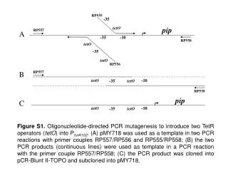
RP555 -35 tetO pip RP557 A -35 -10 RP558 tetO -35 tetO RP556 RP557 B tetO -10 -35 tetO RP558 pip C tetO -10 -35 tetO Figure S1. Oligonucleotide-directed PCR mutagenesis to introduce two TetR operators (tetO) into PfurA102. (A) pMY718 was used as a template in two PCR reactions with primer couples RP557/RP556 and RP555/RP558; (B) the two PCR products (continuous lines) were used as template in a PCR reaction with the primer couple RP557/RP558; (C) the PCR product was cloned into pCR-Blunt II-TOPO and subcloned into pMY718.
HindIII HindIII Pptr lacZ HindIII PfurA102tetO HindIII pip HindIII HindIII PfurA102 Pptr lacZ pip pMY718 HindIII Ligase HindIII PfurA102tetO HindIII Pptr lacZ pip pFRA40 Figure S2.pMY718 was cut with HindIII to remove the PfurA102-containingfragment. The backbone of the plasmid was ligated with the HindIII fragment containing PfurA102tetO. Correct orientation was determined by sequencing the junction between the two fragments.
pip Sm oriE lacZ tetR integrase PfurA102tetO Pptr Psmyc pip Sm oriE lacZ tetR integrase PfurA102tetO Pptr Psmyc pFRA42A pFRA42B Figure S3. Structure of the integrative plasmids pFRA42A and pFRA42B. Bent arrows represent promoters. Pptr: Pip-dependent promoter; Psmyc: constitutive promoter controlling tetR transcription; PfurA102tetO: TetR-dependent promoter controlling expression of pip. The two plasmids differ for the orientation of the Psmyc-tetRcassette.
Hyg pFRA51/53 Pptr ftsZ or fadD32 Hyg Pptr ftsZ,fadD32 Figure S4. Strategy used to replace ftsZ or fadD32 promoters with Pptr by insertional mutagenesis. The 5’-portion of each gene was cloned downstream of Pptr in the suicide vector pFRA50. After integration of the resulting plasmids in the mycobacterial genome, an intact copy of the gene is placed under Pptr transcriptional control, whereas the residual 5’-portion is still downstream of the physiological promoter.
CCTGTCACGCTAGCCAAAGTCAACTGAGATTCCAGAAAAGGGAGTCATATTGTCTAGTGTGCCTGTCACGCTAGCCAAAGTCAACTGAGATTCCAGAAAAGGGAGTCATATTGTCTAGTGTG -35 -10 PfurA102 CCTCCCTATCAGTGATAGATCAACTGATCCCTATCAGTGATAGACATATTGTCTAGTGTG -35 -10 tetO tetO PfurA102tetO Figure S5. Sequence of PfurA102 and PfurA102tetO. The -35 and -10 sequences are underlined, tetO operators are boxed.
MS82 MS83 50 ng/ml ATc 10 ng/ml ATc no ATc A Figure S6. Characterization of the TetR/Pip OFF system. b-galactosidase assay on solid media. M. smegmatis MS82 and MS83 were plated on Middlebrook 7H10 plates containing X-gal and different ATc concentrations.
B A Figure S7. Morphology of the M. smegmatisftsZ conditional mutant MS98. Cultures grown in the absence (A) in the presence (B) of 50 ng/ml ATc were examined by light microscopy with a 100x objective.
Figure S8. Ziehl-Neelsen staining of the M. tuberculosisfadD32 conditional mutant grown in the absence (A) or in the presence (B) of 200 ng/ml ATc. Bacteria were incubated for 72 h, and examined by light microscopy with a 100x objective. Ziehl-Neelsen positive bacteria are stained in red, Ziehl-Neelsen negative bacteria are stained in blue.