Download
1 / 18
180 likes | 271 Views
Learn the detailed structure and function of skeletal muscles, including the contraction process, motor unit innervation, and energy sources involved. Explore muscle fiber types, force summation, and effects of hypertrophy and atrophy.

E N D
Contraction of Skeletal Muscle Arsalan Yousuf BS 4th Semester
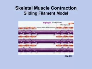
Anatomy of Skeletal Muscles • All skeletal muscles are composed of fibres ranging from 10-80 uM in diameter. • Sarcolemma • Myofibrils (Actin and Myosin filaments) • Each myofibril is composed of : • 1500 myosinfilaments • 3000 actin filaments. • Ends of the actin filaments are attached to Z-disc. • The portion of the myofibril that lies between two successive Z discs is called a sarcomere. • Sarcoplasm and sarcoplasmic reticulum. • The actin and myosin filaments are kept in place by a large number of filamentous molecules of a protein called titin.
Mechanism of Muscle Contraction • Action potential travels along a motor nerve to its endings on muscle fibers. • The nerve secretes neurotransmitter acetylcholine. • Acts on muscle fiber membrane to open multiple “acetylcholine-gated” channels • Allows sodium ions influx into the muscle fiber and initiates action potential at the membrane. • The action potential travels along the muscle fiber membrane in the same way that action potentials travel along nerve fiber membranes. • The action potential causes the sarcoplasmic reticulum to release large quantities of calcium ions that have been stored within this reticulum. • The calcium ions initiate attractive forces between the actin and myosin filaments, causing them to slide alongside each other, which is the contractile process. • After a fraction of a second, the calcium ions are pumped back into the sarcoplasmic reticulum by a Ca2+ membrane pump this removal of calcium ions from the myofibrils causes the muscle contraction to cease.
Myosin Filament • Made up of 200 or more individual myosin molecules having a molecular weight of about 480,000. • The myosin molecule is composed of six polypeptide chains: • two heavy chains (MW 200,000) • four light chains (MW 20,000) • Myosin head functions as an ATPase enzyme to energize the muscle contraction process.
Actin Filament The actin filament composed of three protein components: actin, tropomyosin, and troponin. Troponin are actually complexes of three loosely bound protein subunits (Troponin I, T and C), each of which plays a specific role in controlling muscle contraction.
Molecular Basis of Muscle Contraction • Active sites on the normal actin filament of the relaxed muscle are covered by the troponin-tropomyosin complex. • Troponin complex undergoes conformational upon binding of Ca2+ binding. • ATP as the Source of Energy for Contraction—Chemical Events in the Motion of the Myosin Heads.
Sources of Energy for Muscle Contraction Energy is utilized during: • Walk-along mechanism. • Pumping calcium ions from the sarcoplasm into the sarcoplasmic reticulum. • Pumping sodium and potassium ions through the muscle fiber membrane. Energy comes through • ATPin the muscle fiber is about 4 mM. • Phosphocreatinine (concentration 5 times higher than ATP) • Glycogen • Oxidative metabolism (95% of energy is obtained through this mechanism)
Fast Fibres vs Slow Fibres • The muscles that react rapidly are composed mainly of “fast” fibers. • The muscles that respond slowly but with prolonged contraction are composed mainly of “slow” fibers.
Motor Unit: All the muscle fibers innervated by a single nerve fiber are called a motor unit. small muscles that react rapidly and whose control must be exact have more nerve fibers for fewer muscle fibers Opposite is the case for large muscles e.g. soleus muscles
Force Summation: • Summation means the adding together of individual twitch contractions to increase the intensity of overall muscle contraction. • Can occur in two ways: • Multiple fiber summation • (increase in number of motor • unit contractions) • Frequency summation • (increasing the frequency of • contractions)
Muscle Hypertrophy and Atrophy. • When the total mass of a muscle increases, this is called muscle hypertrophy. When it decreases, the process is called muscle atrophy. • Muscle denervation could lead to atrophy. • When a muscle remains unused for many weeks, the rate of decay of the contractile proteins is more rapid than the rate of replacement.