Download

1 / 14
340 likes | 930 Views
ULTRASOUND GUIDED CENTRAL VENOUS CANNULATION. By Dr Sunil Chhajwani (MD. Anaesthesia). A video presentation. Indications : Haemodynamic monitoring Infusion of inotropes, vasodilators, vasopressors pacing Aspiration of air embolised into right side of heart Infusion of fluids.

E N D
ULTRASOUND GUIDED CENTRAL VENOUS CANNULATION By Dr Sunil Chhajwani (MD. Anaesthesia)
Indications : Haemodynamic monitoring Infusion of inotropes, vasodilators, vasopressors pacing • Aspiration of air embolised into right side of heart • Infusion of fluids
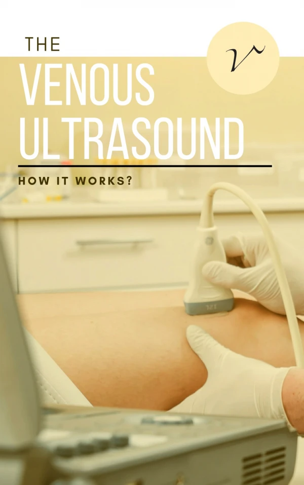
Placement of ultrasound guided central venous catheter • Ultrasound (with high resolution probe)to be kept at the head end of patient • Probe to kept transversely caudad to needle placement • Probe marker should face patient's left side • Trace the IJV from angle of mandible to supraclavicular fossa using linear probe in transverse orientatiion
Assessment of IJV • IJV diameter should be 7 mm. • Avoid access point to IJV where there is overlap with carotid artery • Rule out thrombus in IJV • Avoid head tilt more than 30 degrees to avoid transversing carotid artery
Use local anaesthetics without adrenaline (to prevent inadvertent injection into carotid artery) • CVC insertion site should be prepared with usual sterile technique • Ultrasound gel should be applied to linear probe and sterile cover to be placed over the probe • Make sure no air bubbles between face of probe and sterile sleeve.
IJV should be imagined and placed in centre of ultrasound field • Needle should be angled at 40-60 degrees at the angle of neck and 1 cm back from the middle of ultrasound probe • If the needle is aligned correctly the soft tissue depression should lie exactly over the IJV • Advance the needle in small increments of 0.5 cm
If the needle is seen to grow medially or laterally , it is withdrawn till below skin tissue and then directed towards IJV. • Correct placement of needle is indicated by indent on IJV wall. • Make sure needle is seen inside the IJV lumen • Aspirate free flow of blood from IJV • Pass guide wire through the puncturing needle
Look for guide wire inside the lumen of IJV by USG probe • which is seen as hyper echoic dot like shadow when probe is kept transversely or • hyperechoic straight shadow when probe is kept longitudinally to IJV • Dilate the tract with help of dilator • Pass central venous catheter over guide wire • Confirm the position of cvc by USG and free aspiration of blood from all the lumens.