Download
1 / 50
890 likes | 1.78k Views
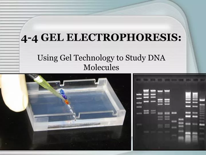
4-4 GEL ELECTROPHORESIS:. Using Gel Technology to Study DNA Molecules. What is Gel Electrophoresis ?. Electro- = flow of electricity -phoresis = to carry across A gel is a suspension of tiny particles in a medium, occurring in a solid form (like gelatin)

E N D
4-4 GEL ELECTROPHORESIS: Using Gel Technology to Study DNA Molecules
What is Gel Electrophoresis? • Electro- = flow of electricity • -phoresis = to carry across • A gel is a suspension of tiny particles in a medium, occurring in a solid form (like gelatin) • Gel electrophoresis= a process that uses electricity to separate charged molecules- DNA, RNA, and proteins, on a gel slab.
Examples • Gel Electrophoresis is used to visualize… • Genomic DNA • RNA • Polypeptides • PCR products • Plasmids • Restriction enzyme digest products http://www.steve.gb.com/images/science/agarose.jpg
Two Types of Gels • Agarose gels • Medium to Large DNA fragments or RNA • Made from polysaccharides found in various marine species of red algae (Agar)— • Some found right here in California! • Agar is composed of both agarose and agaropectin molecules and provides support to the cell walls within the marine algae. Removed from the plant, the agar can be used as a food thickener, much like gelatin, a laxative, or a medium for growing bacteria, fungus, or other microorganisms, when purified.
Two Types of Gels • Agarose gels • The Agarose is purified and dissolved in a boiling buffer solution (TAE) • When solidifies it provides network of pores (a matrix) for particle movement • Run in horizontal gel boxes • Gel concentration depends on the size of the DNA fragment of interest • Most common stain to visualize the DNA: Ethidium bromide (EtBr) or Methylene blue
Two Types of Gels 2) Polyacrylamide Gels (PAGE) • For proteins and very small DNA/RNA fragments (get finer resolution of bands) • Made from the polymer, polyacrylamide (toxic in liquid state; non-toxic in solid state) • Run in vertical gel boxes • Gel concentration depends on the size of the protein of interest • Most common stain to visualize Proteins: Coomassie Blue or Silver Stain
Why Perfom gel electrophoresis? • When DNA is cut by restriction enzymes*, the result is a mixture of DNA segments of varying lengths • It is useful to be able to separate the pieces (for forensic work or for DNA sequencing) *Reminder: …. A few words about restriction enzymes!
History of Restriction Enzymes • Restriction enzymes are called restriction endonucleases- cut DNA strand at certain nucleotide sequences usually 4-6 base pairs long • Occur naturally in some species of bacteria where their role is “defense” • These enzymes “restrict” foreign (e.g. viral) DNA that enters the cell, by destroying it—cutting the invader DNA at certain segments.
The host cell DNA is protected • ~800 known restriction enzymes • 1971- Biotechnology uses restriction enzymes • Restriction Digests – put restriction enzyme with DNA of interest to get known DNA fragments
Restriction Enzymes/Uses: • Lambda DNA (virus that infects bacteria) cut with HindIII—most common molecular weight marker to run as standard on gels • EcoRI (enzyme from E. coli) • BamHI (from Bacillus amyloliquefaciens)
How does gel electrophoresis work? • DNA is an organic macromolecule that has an overall negative charge due to the negative charge on the phosphate groups of the backbone • When the DNA is exposed to an electrical field, the particles migrate toward the positive electrode (hint: think “run red”)
How does it work? • The solution is poured into gel trays with plastic combs. These combs help mold the wells when the hot liquid is poured into the gel tray. When the gel solidifies, wells are left which provide a place to load your DNA. • The solidified gel is placed into a gel box • The gel is covered with TAE buffer and the gel box is filled with the TAE buffer
How does it work? • When the power supply is turned on, an electric current runs into the gel box, and an electric field is established between the positive and negative electrodes. • The charged molecules move out of the wells and into the gel. If the molecule has a negative charge, they migrate towards the positive electrode (DNA). If the molecule has a positive charge, they move toward the negative electrode.
How does it work? • DNA fragments migrate toward the positive electrode at a rate that depends on their size and the size of the pores in the gel. • Large DNA fragments= move slower and stay closer to the wells • Smaller DNA fragments = move faster and move farther from the wells
Different gel [] • The concentration of the gel (matrix density) is critical • The more agarose = the more compact and tighter the matrix = harder for charged particles to move through the gel • Most agarose gels are made between 0.6% and 3%. • A 0.8%gel (most common) will show good resolution (separation) of large DNA fragments (500-10,000 bp)
Different gel [] • A 2%-3% gel will show good resolution for small DNA fragments (200-1,000 bp) • Lower percentage gels (0.6%) are very weak and may break when you try to lift them. They separate only VERY Large DNA = genomic DNA. • High percentage gels are often brittle and do not set evenly.
What is needed? • Solidified Agarose gel • Power supply • Gel box (chamber)
What is needed? • Buffer- either TBE or TAE • The buffer provides ions in solution to ensure electrical conductivity • Contains TRIS, a buffering salt that stabilizes the pH and helps maintain the shape of the molecules being analyzed • Not only is the agarose dissolved in buffer, but the gel slab is submerged in the buffer after adding it to the gel box
What is needed? • Sample Loading Buffer– • To visualize sample movement through the gel (you ARE NOT visualizing the DNA as it moves through the gel!) • We cannot “see” the DNA until we stain it • Helps “sink” the DNA into the wells (glycerol or equivalent) • The loading dye helps us keep track of how far the DNA has migrated through the gel • You don’t want your DNA to run off the gel!
What is needed? • Molecular weight markers (Ladders)- DNA fragments of known size (Lambda/HindIII) • Comparison of your sample bands to the marker bands allows… • Visible confirmation of desired product • Quantification of sample DNA http://www.biomedcentral.com/content/figures/1471-2180-5-63-2.jpg
Gel electrophoresis DNA fragments - electrode + electrode Agarose gel ~~~~~~~~~~~~~~~~~~~~~~~~ ~~~~~~~~~~~~~~~~~~~~~~~~ buffer ~~~~~~~~~~~~~~~~~~~~~~~~ ~~~~~~~~~~~~~~~~~~~~~~~~
Gel electrophoresis - electrode + electrode current ~~~~~~~~~~~~~~~~~~~~~~~~ ~~~~~~~~~~~~~~~~~~~~~~~~ buffer ~~~~~~~~~~~~~~~~~~~~~~~~ ~~~~~~~~~~~~~~~~~~~~~~~~
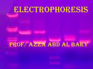
Visualizing DNA • Most of the time Ethidium bromide (EtBr) is used • A fluorescent dye visualized when excited by UV light • Intercalates (inserts) into the DNA molecule, thereby “staining” it
Visualizing DNA • Gel is soaked in a solution of EtBr and the DNA bands take up the dye (or EtBr is added directly to the gel solution during preparation) • Then the gel is then placed on a light box, exposed to UV light, and a photograph is taken for documentation
Brighter bands= high concentration of DNA Lighter bands = low concentration of DNA **Each band that you see is a collection of millions of DNA molecules, all of the same length!!
Visualizing DNA • Ethidium Bromide is a mutagen • So we will use trace amounts of EtBr in our gelstominimize student exposure to the compound • Listen to instructions when pouring your gels or analyzing your gels! (CMB)
Important! • A gel only shows us the relative size of the DNA fragments. It DOES NOT tell us the sequence of those fragments • DNA sequencing is the next step to determining the actual nucleotide sequence of the DNA fragment(s).
Gel Electrophoresis Prepare agarose gel Melt, cool and add Ethidium Bromide. Mix thoroughly. Pour into casting tray with comb and allow to solidify Add running buffer, load samples and marker Run gel at constant voltage until band separation occurs View DNA on UV light box and document results
Helpful Hints • When placing the gel in the electrophoresis chamber: • Make sure that the wells are closest to the negative (black) electrode (since DNA is negative) • When adding the buffer to the chamber: • Gently flood the gel from the end opposite the wells to minimize sample diffusion • Before loading the wells: • Orient the entire chamber close to the power supply so it is in place when the samples are ready to run
Helpful Hints • When loading samples in the wells of the gel: • Use proper micropipette techniques • Make a written record of which sample you will load in each well of the gel • Be careful not to puncture the bottoms of the wells as you load the samples • If you make a mistake, do NOT take more than the allotted sample; there are limited amounts
Troubleshooting Guide DNA Science: A First Course in Recombinant DNA Technology, by David A. Micklos and Greg A. Freyer
Gel electrophoresis separates molecular products based on: Shape Size Charge Evolutionary similarity
Why does DNA move through a gel? • The negative charge of DNA is attracted to the positive end of the electrophoresis box. • The negative charge of DNA is attracted to the negative end of the electrophoresis box. • The positive charge of DNA is attracted to the positive end of the electrophoresis box. • The positive charge of DNA is attracted to the negative end of the electrophoresis box.
What is the purpose of loading dye in gel electrophoresis? • To see the DNA in the gel • To help your sample sink into the well • To “see” movement on the gel so you can turn it off before any DNA runs off the edge of the gel • Both 2 & 3
What is the purpose of a DNA ladder? • So you can estimate the length of DNA fragments running through a gel • It’s a control for your experiment • It helps DNA samples move through the gel • Both 1 & 3
When traveling through an agarose gel, larger molecular products will migrate _________ smaller molecular products. Faster than Slower than At the same rate
Which fragment is the largest? • Fragment #1 • Fragment #2 • Fragment #3 • Fragment #4 1 2 3 4
The size of the molecular products is determined by: The intensity of the band The percentage of agarose gel Comparison with a molecular weight “ladder” Being familiar with your gel and “eyeballing” it
The concentration of the molecular products is determined by: The intensity of the band The percentage of agarose gel Comparison with a molecular weight “ladder” Being familiar with your gel and “eyeballing” it
Buffer is used instead of water when making and running gels because: Buffer enhances the transmission of electric currents in water Buffer is more homogeneous than water Buffers provide the salts necessary to help create the current
How Gel Electrophoresis Works http://www.sumanasinc.com/webcontent/anisamples/majorsbiology/gelelectrophoresis.html http://learn.genetics.utah.edu/units/biotech/gel
If this is a P200, what volume will be measured? 1000uL 100uL 10uL 1uL
If this is a P1000, what volume will be measured? 0 3 0 3000uL 300uL 30uL 3uL
If this is a P20, what volume will be measured? 1 7 3 1730uL 173uL 17.3uL 1.73uL
What is the purpose of the 1st stop on a micropipettor? Measure correct volume Eject entire volume from the tip Eject the tip from the pipette None of the above
Is this centrifuge balanced? yes no