Download
1 / 61
980 likes | 2.11k Views
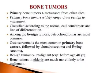
Pazourek L., Ondrůšek Š. Malignant bone tumors. Osteosarcoma. Malignant osteoid. Epidemiology. 3 new cases /1 milion/ year 2. decade Metaphysis of long bones 1/2 in knee region distal femur proximal tibia proximal humerus. Classification. Primary Central High-grade

E N D
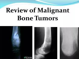
Pazourek L., Ondrůšek Š. Malignant bone tumors
Osteosarcoma Malignant osteoid
Epidemiology • 3 new cases /1 milion/ year • 2. decade • Metaphysis of long bones 1/2 in knee region distal femur proximal tibia proximal humerus
Classification • Primary • Central • High-grade • Conventional high-grade (80 – 90%) • Osteoblastic • Chondroblastic • Fibroblastic • Telangiectatic • Low-grade
Peripheral • High-grade • Low-Grade • Parosteal (low-grade) • Low/intermediate-grade • Periostal
Secondary - in Paget´s disease of bone - post radiation
Symptoms • pain • during night, in rest • swelling • pathological fracture • metastases in the time of diagnosis in 10-25 % of patients
Diagnostics • X-ray • CT / MRI • Scintigraphy • Chest X- ray or spiral CT • Ultrasonography • Biopsy – excisional, needle
OSA • Missed case • Wrong prognosis Oncologic reflex
Therapy • neodjuvant chemotherapy • surgery – radical resection / amputation • adjuvant chemotherapy • Metastasectomy in lungs • Chemotherapy: (EURAMOS protocol) • metotrexat, doxorubicin, adriamycin, cisplatina, ifosfamid, etoposid. • In low-grade OSA – only surgical treatment • OSA is a radioresistant tumor
Prognostic factors • Metastases • Size of the tumor • Axial localisation • Radicality of surgery • Response to chemotherapy
Prognosis – 5 years survival • 70% - conventional high-grade OSA without MTS and with good respone to chemotherapy (to 10 % of vital tumor cells) • 90% - u low-grade OSA after radical surgery
Epidemiology • 10% of primary malignant bone tumors • Age: • primary: 40 – 60 years • secondary: 25 – 45 years • Localisation- pelvis, proximal femur, proximal humerus
Etiology • Secondary • Multiple enchondromas (M.Ollier, Maffucci sy • Exostosis disease cartilage over 2 cm • Chondroblastoma, chondromyxoid fibroma …
Therapy • Radical resection – wide resection, amputation • Metastasectomy in lungs • Chemoresistant tumor • Radioresistant tumor
Prognosis • Prognostic factors: • Radicality of surgery • Size • Histological grading • In intralesional surgery – 90% risk of local recurrence and lung metastases • Prognosis: • Conventional low-grade 90% 10 years • Conventional high-grade 20-40% 10 years • Dediferenciated sarcoma 15% 5 years
Ewing sarcoma family • Group of high grade malignant round cells bone tumors with neuroectodermal differentiation and specific translocation. • Ewing sarcoma • PNET (periferal neuroectodermal tumor • Askin tumor of the chest wall • Neuroblastoma in adults
Epidemiology • One new case /1 mil./ 1 year • 5-25 years • In metaphysis of long bones with extension into diaphysis and in flat bones (pelvis, scapulla)
Symptoms • pain • swelling • Fever, redness, • Leucocytosis, ESR elev. • Biopsy- + identification of specific gene translocation t(11,22)q(24,12)
Therapy • Chemo and radio sensitive tumor • Neoadjuvant chemotherapy • Local therapy: • Radiotherapy • Wide resection • Radiotherapy and wide resection • Adjuvant chemotherapy • In risk patients: transplantation of bone marow • Metastasectomy in lungs
Prognosis • Response to chemotherapy (systemic disease) • 5-years survival in 60 % of patients • Worse prognosis: • metastases • Size over 100cm3 • Surgery not possible • Axial localisation • Local recurrence • Some genetic variants
Malignant fibrous histiocytomain bone • In 5. decade • In long bones – femur, tibia • X- ray osteolytic lesion + cortical erosions, soft tissue mass • Therapy: neoadjuvant chemotherapy + wide resection or amputation + adjuvant chemotherapy • It is a radioresistant tumor • Survival 35 % 5 years
Adamantinoma • Very rare • 90 % in tibia • Therapy: radical resection • Radioresistant tumor • Prognosis – unclear
Chordoma • Axial localisation • Osteolytic lesion • Th- radical surgery or radiotherapy • Prognosis- bad
Malignant vascular tumors • Hemangioendotelioma • Hemangiopericytoma • Angiosarcoma • Osteolytic lesions • Therapy: wide resection or amputation • Chemotherapy in high grade • Radiotherapy in non oper. cases
Primary bone tumors • Multiple myeloma (plasmocytoma) • Solitary plasmocytom (myelom) • Primary bone lymfoma • Secondary lesions • Hodgkin lymfoma • Non-Hodgkin lymfoma • Leukemia Therapy- chemotherapy and radiotherapy in hematooncology
Multiple myeloma • Most often bone tumor (40%) • 5 – 6. decade • Symptoms: • pain • Pathological fracture • weaknes • letargy • infections • Renal failure • Headache
Solitary plasmocytoma • Rare • Osteolytic lesion • Resection with replacement + chemotherapy • Prognosis- better than in multiple myeloma
Carcinoma with MTS into the skeleton • Breast • Prostate • Lung • Kidney • Thyreoidal gland
Localisation • Axial skeleton, pelvis, ribs, proximal femur and humerus X-ray • Osteolysis, osteosclerosis, periostal reaction