Download
1 / 74
740 likes | 930 Views
Learn about the structure of the human body through macroscopic and microscopic anatomy, embryology, and directional terms. Explore axes, planes, and divisions of body cavities to gain a comprehensive understanding. Access e-learning resources and lectures for a better grasp of anatomical concepts.

E N D
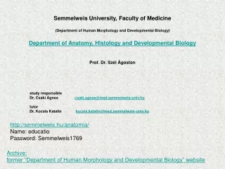
Semmelweis University, Faculty of Medicine Anatomyforstudents of Faculty of Pharmacy (GYTK) (formerDepartmentof Human Morphology and DevelopmentalBiology) Department of Anatomy, Histology and Embryology head of theDepartment: Prof. Dr. Szél Ágoston lecturer Dr. Csáki Ágnes csaki.agnes@med.semmelweis-univ.hu tutor Dr. Kocsis Katalin kocsis.katalin@med.semmelweis-univ.hu itc.semmelweis.hu e-learning / Moodle SeKA login name and password histologyslidesvia internet http://semmelweis.hu/anatomia/ Name: educatio Password: Semmelweis1769
onlytheanatomicalpages (seetheexamrequirements) Home page: http://semmelweis.hu/anatomia Lectures Facultyof Pharmacy (GYTK) Anatomy
STRUCTURE OF THE HUMAN BODY INTRODUCTION Anatomydealswiththestructure of the body. MacrosopicAnatomy: studiescomponents, whichcan be seenwithunaidedeye MicroscopicAnatomy (Histology): examinestheinvisiblecomponentssuchasdifferenttissues, cells building upthe human body. Light and electronmicroscopesareusedtostudythesecomponents. Embryology: development of theembryo
Axes, planes and directionshelptodetermine theposition and relation of thestructuralcompartments of the body toeachother. Anatomicalposition:erected human body, with palmfacesanteriorly New „language”, need „vocabulary” The principalaxes and planes of the body
Principalaxes of the body 1.LONGITUDINAL (VERTICAL) AXIS:Y longaxis of the body is verticalwhen the body is heldin an uprightpositon 2. TRANSVERSE (HORIZONTAL) AXIS:X It is perpendiculartothelongitudinalaxisfrom right toleft 3. SAGITTAL AXIS: Z runsfromthe back tothe front surface of the body like an arrow, it is perpendicularto theabove-mentionedtwoaxes
Principalplanes of the body 1. MEDIAN SAGITTAL PLANE: The imaginaryverticalplanethatdividesthe body longitudinallyintoexact right and leftsymmetricalhalves. SAGITTAL PLANEs: Anyplanewhich is parallel tothemedianplane. 2. FRONTAL (CORONAL) PLANE: Verticalplanedividesthe body intoanterior and posteriorparts. Itcontains (runthrough) thetransverse and longitudinalaxes, laying parallel totheforehead and perpendiculartothesagittalplanes. 3. TRANSVERSE PLANE: It is horizontalintheuprightposture, containsthesagittal and transverseaxes, liesperpendiculartothesagittal and coronalplanes.
DIRECTIONS CRANIAL: towardthehead (cranium) CAUDAL: towardthebuttocks (cauda: tail) SUPERIOR (ABOVE): aboveortowardthehead INFERIOR (BELOW): beloworawayfromthehead DEXTER: right side SINISTER: leftside MEDIAL (CLOSER TO THE MIDLINE): relatingtothemiddleor center LATERAL (AWAY FROM THE MIDLINE): towardstheside of the body ANTERIOR: toward the front POSTERIOR: toward the back VENTRAL: toward the abdomen (venter: stomach) DORSAL: toward the back (dorsum) SUPERFICIAL (PERIPHERAL): toward the body surface PROFUNDUS (DEEP): toward the inner of thebody EXTERNAL: outside INTERNAL: inside
DIRECTIONS ON THE EXTREMITIES PROXIMAL (closertothepoint ofattachment): towardthetrunk DISTAL (away from the point of attachment): away from the trunk PALMAR (volar): on or toward the palm of the hand PLANTAR: on or toward the sole of the foot ULNAR: toward the ulna RADIAL: toward the radius TIBIAL: toward the tibia FIBULAR: toward the fibula
CAPUT (HEAD) • COLLUM(NECK • TRUNCUS (TRUNK) • UPPERLIMB • LOWERLIMB DIVISIONS OF THE BODY
CAVITIES OF THE HUMAN BODY I. CRANIAL CAVITY Houses the brain II. THORACIC CAVITY Houses the heart, main vessels, lungs, esophagus, trachea III. ABDOMINAL CAVITY Contains the stomach, intestines, liver, kidneys, pancreas. THORACIC DIAPHRAGM Separates the thoracic cavity from the abdominal ones PELVIS containsurogenitalorgans and rectum PELVIC DIAPHRAGM Flooring, closestheabdominalcavity, separetesthepelvisfromoutergenitals
it is an innerframework, providessupport and structuretothe body, maintainsour body shape • provides a system of muscleleversthatallow body movement • containsbonemarrow, thebloodformingtissues of the body • borders cavities, protectstheinnerorgans • a human skeleton is generallyformedbyabout 206 separatebones. • bonesareconnectedtootherbonesbyjoints Axialskeleton – skull, vertebralcolumn (spine), ribs, sternum Appendicularskeleton - limbs LocomotorsystemSKELETON
Structure of the bones 1.COMPACT (CORTICAL) BONES: - Hard and dense, observedonthesurface • Consist of Haversiansystems 2.TRABECULAR (SPONGY) BONES: • Fillstheinterior of bones • Composed of spongy-likenetwork • Best adaptedtoresist of local strains • Spaceforbloodvessels and bonemarrow Spongy-like material Compactbone
Compact: Organized into concentric units called osteons or Haversian systems. Each osteon contains a single, central canal called the Haversian canal and concentric layers of hardened matrix, called lamellae; canals that extend between the osteonic canals called Volkmann’s canalsforvessels Spongy bone: consists of numerous bony plates with spaces in between filled with red marrow the cells are embedded within the lacunae in the bony plates. (trabeculae)
Shape of bones: • longbones (e.g. humerus) • flatbones(e.g. scapula, skull) • shortbones (e.g. wrist, ankle) • irregularbones (e.g. vertebrae, sacrum) • pneumaticbones (viscerocranium)
Ontheoutersurface of bones Twolayers: outerfibrous and innerosteogenic, cellularlayer Osteogeniclayer – thiknessgrowing of bone, repair of fracture Bloodsupply, nervesupply of bone Bone marrow ADDITIONAL COMPARTMENTS OF BONEPeriosteum Red bonemarrow: - Haemopoeticactivity (production of bloodcells) Fetty (yellow) bonemarrow: - no haemopoeticactivity
The individual bones of the skeleton are connected either continuously or discontinuously. CONTINUOUS JOINTS (SYNARTHROSES) relativelyimmobilejointswhereindifferenttypes of rigidconnectingtissuesjointwobones thesejointsprovideskeletalstabilityatthejunction of thebones Types of continuous joint 1. FIBROUS CONNECTION(SYNDESMOSIS) 2. CARTILAGINEOUSCONNECTION(SYNCHONDROSIS) 3. BONYCONNECTION (SYNOSTOSIS)
is the most common and movabletype of joints Components of the synovial joints obligatory 1. articular surfaces 2. articularcartilage 3.joint capsule 4.jointcavity 5. ligaments additional articular diskormeniscus, articularlip, DISCONTINUOUSORSYNOVIAL JOINTS (ARTICULATIONES)
Articular surfaces bony socket (cavum) concave part of a joint bony head (caput) end of the bone fitting into the bony socket Articular cartilage hyalinecartilageorfibrocartilage function is toabsorbshock and reducefrictionduringmovement Jointcapsule surrounds and unitesthearticulatingbones, consistsof two layers (1) theouterfibrousmembrane (2) theinnersynovialmembranethatsecretesthelubricating, and joint-nourishingsynovial fluid Jointcavity Spacebetweenthebonesthat is filledwithsynovial fluid Ligaments dense, regular connective tissue highlyadaptedforresistingstrainstopreventextrememovementsthatmaydamagethejoint
Disc and menisc The articular disc completelyseparates thearticular cavity into two compartments Ameniscus is an incompleteC-shapedfibrocartilagebetween the two bones of knee that serves as a shock absorber. These can often be injured in sports or traumatic injuries, but they can also be torn and damaged with normal wear and tear activities as we mature. There are actually two meniscal discs, one on the medial side and another on the lateral side of theknee joint
TYPES OF JOINTS MULTIAXIAL JOINTS BALL AND SOCKET JOINT BIAXIAL JOINTS ELLIPSOID (condyloid) JOINT SADDLE JOINT UNIAXIAL JOINTS HINGE JOINT PIVOT JOINT
Movement possibilities of the joints Abduction Adduction Aroundsagittalaxis: Abduction: movementawayfromthemidline of the body Adduction: movementtowardthemidline of the body Aroundtransverseaxis: Extension: straighteninglimbsat a joint , and Flexion: bendingthelimbsat a joint Aroundlongitudinalaxis: Rotation: a circularmovement Circumduction: combined, circular movement Flexion Extension Specialmovements Opposition - reposition - thumb Eversion - inversion - foot Protraction - rettraction - head Elevation - depression - shoulder Pronation- supination - forearm Circumduction Rotation
Ball and socket joint (multiaxial joint) the bony head is rounded the bony socket is a cuplikesurface examplesaretheshoulder and the hipjoint Movements: flexion-extension adduction-abduction rotation circumduction
Saddle (sellar) joint (biaxial) consists of two saddle shaped articular surfaces each having a concave and a convex curvature Onlyoneexample: the carpometacarpal joint of the thumb Movements: Opposition-reposition, circumduction
Ellipsoid joint (biaxial) the oval-shaped end bone fits into an oval-shaped cavity (eggin an egg-cup), e.g. wrist joint Movements: flexion-extension adduction-abduction circumduction
Hinge joint (ginglymus) (uniaxial) the convex articular surface fits into a groove of the concaveone example: knee and ankle joint Movement: flexion-extension
Pivot (trochoid) joint (uniaxial) the convex articular surface fits into a groove of the concaveone allowsrotationarroundlongitudinalaxis Intheforearmstheradius and ulna twist arroundeachother inthenecktheskull and thefirstvertebraspins over thesecondcervicalvertebra (C1-C2) Movement: Rotation
Plane (gliding) joint permitsslidingorglidingmovementsintheplane of articularsurfaces the opposedsurfaces of thebonesareflator almost flat the movement limited bytheirtightjointcapsules Example: intertarsal, intercarpaljoints
Tendon Origin Muscle belly Tendon Insertion The bones and the joints represent the passive part of the locomotor system. The skeletal muscles form the active part of it. Myology is thedescription of muscles. Skeletal muscle Skeletalmuscle is undercontrol of thesomaticnervoussystem Origin of the skeletal muscle is (usually)ona less mobile bone Insertion (attachment) is (usually)ona more mobile bone
Forms of the sceletalmusclesbytheorigin two-headed muscles (biceps) three-headed muscles (triceps) four-headed muscles (quadriceps)
Types of the muscles: fusiform (spindleshaped) muscles long fibers,produce extensive movements flat muscles (convergent) flat tendon or aponeurosisforattachement unipennate muscles long tendon, musclefibersonlyononeside bipennate muscles(„feather”) the muscle fibers attach to both sides of the tendon sphincters circularmuscles maintainsconstriction of a natural body passageororifice relaxesasrequiredbynormalphysiologicalfunctioning
Types of the muscles with oblique fibers unipennate muscles long tendon, musclefibersonlyononeside bipennate muscles(„feather”) the muscle fibers attach to both sides of the tendon
FLEXORS (e.g.biceps on the shoulder joints) EXTENSORS (e.g.triceps on the shoulder joint) Ú ADDUCTORS (e.g.adductors of the thigh) Classificationaccording to their functions
Synergistic (agonist) muscles groupsof musclesthatcontracttogethertoaccomplishthesame body movement Example:biceps and brachioradialis. The biceps is theprimarymover in elbowjointmovementand thebrachioradialisactsas a synergisticmuscletostabilizethejoint, thusaiding in themotion Antagonistic muscles A musclethatacts in oppositiontothespecificmovementgeneratedbytheagonist and is responsibleforreturning a limbtoitsinitialposition An example of thiskind of musclepairing is thebicepsbrachii and tricepsbrachii. Whenthebicepsarecontracting, thetricepsarerelaxed, and stretches back toitsoriginalposition. The oppositehappenswhenthetricepscontract.
tendon fascia tendon sheath synovial bursa sesamoid bone fatty body Auxiliary features of the muscles
Tendon toughband of fibrousconnectivetissuethatusuallyconnectsmuscletobone buildup of collagenfibers tendons and musclesworktogetherandcanonlyexert a pullingforce Tendon (synovial) sheath Twolayers: Inner or synovial layer secreting the synovial fluids Outer or fibrous layer The two layers glide on each others, lubricated by the synovial fluid
Thoracolumbal Fascia FASCIA - connectivetissuethatsurroundsmuscles, bloodvessels, and nerves, bindingthosestructurestogetherinmuchthesamemannerasa plasticwrap - passivestructuresthattransmitmechanicaltensiongeneratedbymuscular activitiesorexternalforcesthroughoutthebody FASCIA
Synovial bursa smallfluid-filledsaclinedbysynovial membrane provides a cushionbetweenbones and tendons and/ormusclesaround a joint helpstoreducefrictionbetweenthe bones and allows free movement bursaearefilledwithsynovialfluid and arefoundaround most major joints of the body
SKULL, VERTEBRALCOLUMN HEAD, NECKMUSCLES
Skull (cranium) framework of the head the human skull is usually made up of 22 bones protectsthebrain and holdstheotherparts of thehead(eyes, nose, mouth, etc.) • neurocranium: houses the brain • viscerocranium: facial portion (mouth, nose)