Download
1 / 41
580 likes | 1.51k Views
MUSCLE ORIGIN, INSERTION, AND ACTION. THE MUSCLULAR SYSTEM. Major Skeletal Muscles: Anterior View. The 40 superficial muscles here are divided into 10 regional areas of the body. Figure 10.4b. Major Skeletal Muscles: Posterior View.

E N D
MUSCLE ORIGIN, INSERTION, AND ACTION THE MUSCLULAR SYSTEM
Major Skeletal Muscles: Anterior View • The 40 superficial muscles here are divided into 10 regional areas of the body Figure 10.4b
Major Skeletal Muscles: Posterior View • The 27 superficial muscles here are divided into seven regional areas of the body Figure 10.5b
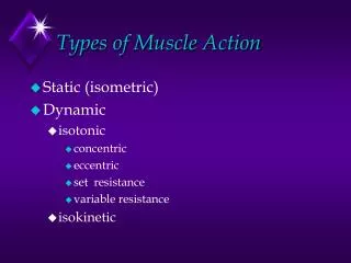
Muscles: Name, and Action • Name and description of the muscle – be alert to information about the muscle given in the name • Origin and insertion – there is always a joint between the origin and insertion • Action– best learned by acting out a muscle’s movement on one’s own body
Muscles of Mastication Figure 10.7a
Frontalis: elevate eyebrows Orbicularis Oculi: close eyelid Zygomaticus: draw angle of lip upward Buccinator: draws cheeks against teeth Orbicularis Oris: closes mouth Platysma: draws lower lip down & back Cranial Aponeurosis: connects frontalis to occipitalis Temporalis: elevates mandible Occipitalis: draws scalp back Masseter: elevates mandible Sternocleidomastoid: Flexes head Draws head toward shoulder Head & Neck Muscles
Muscles of the Anterior Neck and Throat Figure 10.8a
Deeper Muscles of the Neck: Anterior Figure 10.9a
Deeper Muscles of the Neck: Posterior Figure 10.9b
Deep Back Muscles Figure 10.9d
Muscles of Respiration • The primary function of deep thoracic muscles is to promote movement for breathing • External intercostals – more superficial layer that lifts the rib cage and increases thoracic volume to allow inspiration Figure 10.10a
Muscles of Respiration • Internal intercostals – deeper layer that aids in forced expiration • Diaphragm – most important muscle in inspiration Figure 10.10a
Muscles of Respiration: The Diaphragm Figure 10.10b
Muscles of the Abdominal Wall Figure 10.11a
Muscles of the Abdominal Wall Figure 10.11b
Muscles of the Abdominal Wall Figure 10.11c
Extrinsic Shoulder Muscles Figure 10.13a
Extrinsic Shoulder Muscles Figure 10.13b
Muscles Crossing the Shoulder Figure 10.14a
Muscles Crossing the Shoulder Figure 10.14d
Muscles Crossing the Shoulder Figure 10.14c
Muscles Crossing the Elbow Forearm extension • The triceps brachii is the prime mover of forearm extension Forearm flexion • Brachialis and biceps brachii are the chief forearm flexors • The brachioradialis acts as a synergist and helps stabilize the elbow
Forearm: SuperficialAnterior Compartment • These muscles are primarily flexors of the wrist and fingers and pronators Figure 10.15a
Forearm: Deeper Anterior Compartment Deep Deepest Figure 10.15b, c
Forearm:SuperficialPosteriorCompartment • These muscles are primarily extensors of the wrist and fingers Figure 10.16a
Forearm:DeepPosteriorCompartment • These muscles are primarily extensors of the wrist and fingers and the supinator Figure 10.16b
Muscles Crossing Hip and Knee Joints Anterior compartment (most) muscles of the hip and thigh flex the femur at the hip and extend the leg at the knee • Extend the leg (anterior compartment) Posterior compartment muscles of the hip and thigh extend the thigh and flex the leg • Flex and extend the thigh (posterior compartment) Medial compartment muscles all adduct the thigh • Adduct the thigh (medial compartment) • These three groups are enclosed by the fascia lata
Movements of the Thigh at the Hip: Flexion and Extension • The most important thigh flexors are the iliopsoas (prime mover), tensor fasciae latae, and rectus femoris • The medially located adductor muscles and sartorius assist in flexion
Movements of the Thigh at the Hip: Flexion and Extension • Thigh extension is primarily effected by the hamstrings (biceps femoris, semitendinosus, and semimembranosus) • Forceful extension is aided by the gluteus maximus
Movements of the Thigh at the Hip: Other Movements • Abduction and rotation are effected by the gluteus medius and gluteus minimus, and are antagonized by the lateral rotators • Thigh adduction is the role of five adductor muscles (adductor magnus, adductor longus, and adductor brevis; the pectineus, and the gracilis)
Movements:Knee Joint • sole extensor of the knee • quadriceps femoris • flex the knee, and are antagonists to the quadriceps femoris • hamstrings Figure 10.19a
Muscles of the Anterior Compartment primary toe extensors and ankle dorsiflexors • tibialis anterior • extensor digitorum longus • extensor hallucis longus • fibularis (peroneus) tertius Figure 10.21a
Muscles of the Anterior Compartment ISOLATED Figure 10.21b-d
Muscles of theLateral Compartment plantar flex and evert the foot • fibularis longus • fibularis brevis Figure 10.22a
Muscles of the Posterior Compartment primarily flex the foot and the toes • gastrocnemius • soleus • tibialis posterior • flexor digitorum longus • flexor hallucis longus Figure 10.23a
Muscles of the Posterior Compartment - DEEP Deep Figure 10.23b, c
Muscles of the Posterior Compartment - DEEPEST * Deepest Figure 10.23b, c