Download
1 / 29
290 likes | 330 Views
Discover the intricate details of the pelvic girdle and lower limb anatomy crucial for maintaining correct weight bearing and standing postures. Learn about the structure, alignment, and key muscles involved in hip, knee, and ankle movements. Dive deep into the importance of pelvic alignment and muscle flexibility for overall posture and well-being. Explore common conditions like plantar fasciitis and practices like Paschimottanasana to enhance flexibility and release tightness in the lower limb muscles.

E N D
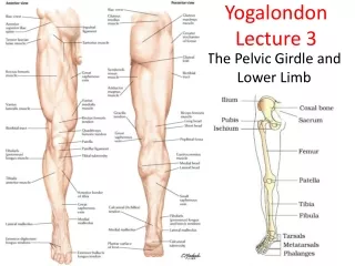
Yogalondon Lecture 3 The Pelvic Girdle and Lower Limb
Pelvic Girdle • The pelvis is formed by 2 innominate bones that meet in the anterior aspect to form the symphysis pubis. • The 2 innominate bones meet the sacrum in the posterior aspect to form the sacroilliac joints(x2) • These articulations form a ring of bone to create a bowl for the internal organs for protection. • Pelvic alignment is vital for correct weight bearing and standing postures.
Pelvis and Hip • Each innominate bone is formed by 3 fused bones, ilium (ASIS, sticky out bits), ischium (sitting bones) and pubis. • The socket in the pelvis is called the acetabulum and together with the proximal end of the femur creates the hip joint.
The Hip Joint • Ball and Socket Joint. (Femur and Acetabulum) - Articulates in the hip socket, with slight inward curve that aligns the body vertically with the knee and ankle. This is fundamental to the centre of gravity and key to standing posture. - Stabilised by very strong ligaments Movements: Flexion, Extension, Abduction, Adduction, Internal Rotation External Rotation.
The Pelvis • The female pelvis is wider, for child bearing and has a definite pubic arch. • As the pelvis is wider in women it could be argued that a wider stance eases standing poses. • Male pelvis is heavier and stronger than female. ?
Muscles of the Pelvic Girdle • Iliopsoas • Extends from the lower back, interior pelvis and attach to femur • Flexor of the hip • What does iliopsoas tightness limit ?
Piriformis • Lies deep and extends from sacrum to femur. • Laterally/externally rotates the hip. Importance of this muscle ?
Muscles of the upper leg • Hamstrings: found at the back of the leg (3parts) • Originate at the ischium of the pelvis insert on the tibia. • Flexes the knee, extends the hip, and externally rotate the knee. • Tightness in hamstrings can affect spine curvature. • Quadriceps: found on front of thigh ( 4 parts) • The 4 parts of the quadriceps combine to form the patellofemoral tendon. • Vastus group: extend the knee • Rectus femoris: extends knee and flexes hip • Rectus Femoris: • Part of the quadriceps muscle on the front of the thigh. Flexes the hip and also extends the knee. 2 joint muscle
Hip Abductors and Adductors • Tensor fascia latae/ ITB • Extends from ilium to ITB and then to tibia • Flexes, internally rotates and abducts the hip • Adductors ( +Sartorius, +Gracilis) • The Adductor muscle group consists of 3 muscles • Extends from the ischium to the femur • Adduct, externally rotate and extends the hip • Gluteus Group (Maximus/Medius/Minimus) • Extends from ilium to femur and into the ITB (iliotibial band) • Extends and externally rotate the hip
The Knee Joint 3x Bones – 2x Joints • Largest joint in the body • Hinge joint (complex) Condylar • Synovial Joint • Articulation between the distal end of femur and tibia. • Articular surfaces covered with cartilaginous discs called meniscus. (medial and lateral) • Stabilised by strong ligaments. • Medial and Lateral collateral ligaments, • Anterior and Posterior Cruciate Ligaments. • The patella forms the patellofemoral joint.
Muscles of the Lower Leg • Gastrocnemius • gives shape to the back of the lower leg and forms Achilles tendon. • Plantaflexes the ankle and assists in flexion of the knee. ( Standing on tip toes ) • Soleus • lies underneath gastrocnemius. • Plantaflexes the ankle, works to keep upright posture. • Tibialis Anterior • Dorsiflexes the ankle.
Ankle and Foot • Ankle: Hinge Joint • 7 tarsus bones. • Foot: 5 metatarsals. 14 phalanges to create toes • The ankle and foot provide the body with both a mobile platform & rigid lever • Movement: Dorsiflexion, Plantaflexion, • Combined movement with the foot producing: Supination & Pronation (inversion & eversion).
Plantar Arch of the foot • Arches of the foot are maintained by • boney structure • plantar muscles • ligaments. • Helps support the body weight. • Weakness or fatigue of muscles can alter arch and foot posture ( flat feet). High arches can be caused by muscular and fascia tightness. • Plantar fascia covers sole of foot and contributes to the formation of the arches.
Plantar fasciitis • Common form of heel pain. • Can be caused by overuse, walking on uneven surfaces, tight calf muscles or over pronation of the foot. • Not an inflammatory condition as once thought but a degeneration of collagen fibres in the plantar fascia. • Improves with stretching.
Paschimottanasana • Lower Limb • Hip: Flexion and adduction • Knee: Extension • Ankle: Dorsiflexion • Passive stretching of hamstrings. Tightness in back of leg and pelvis can cause restriction.
UttanasanaStanding Forward Bend Is there a difference to Paschiamottanasana ?
Setu BandhasanaBridge Pose • Lower Limb • hip extension, adduction, and internal rotation • knee flexion • ankle dorsiflexion • Upper Limb • scapula adduction, downward rotation, elevation; • glenohumeral joint external rotation, extension, adduction; • elbow flexion; forearm supination; • wrist extension (dorsiflexion).
reading for next session 4 • Pages 4, 5 and 10 anatomy colouring book • Pages 23-44 in yoga anatomy edition 2 • Revise for 12 question mini test on lower limb.