Download
1 / 19
230 likes | 473 Views
Senses: Vision. Ch. 17-2. Accessory Structures of Eye. Eyelids Eyelashes Eyebrows Lacrimal apparatus Extrinsic eye muscles. Accessory Structures of Eye. Eyelids – palpebrae Functions: Shade eyes during sleep Protect eyes from light Protect eyes from foreign objects

E N D
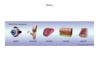
Senses: Vision Ch. 17-2
Accessory Structures of Eye • Eyelids • Eyelashes • Eyebrows • Lacrimal apparatus • Extrinsic eye muscles
Accessory Structures of Eye • Eyelids – palpebrae • Functions: • Shade eyes during sleep • Protect eyes from light • Protect eyes from foreign objects • Spread lubrication over eyeballs • upper eyelid is more moveable – levatorpalpebraesuperioris muscle • Where eyelids meet is called commissure – lateral and medial • Lacrimalcaruncle – reddish bump in medial commissure that secretes oil and sweat – causes “sleep” in your eyes
Infections • Sty – infection in oil gland of eyelash follicle • Chalazion – infection in tarsal glands of eyelid • Tarsal glands secrete fluid onto eye for lubrication
Accessory Structures of Eye • Lacrimal apparatus • Produces and drains lacrimal fluid or tears • Eye muscles
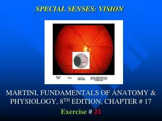
Anatomy of the Eyeball • 2.5 cm in diameter • 3 anatomical layers • Fibrous tunic • Vascular tunic • Retina
Tunic Layers • Outer layer – fibrous • Cornea – transparent, focuses light rays • Sclera – white of the eye • Optic nerve – transmits info to brain • Middle layer – vascular • Choroid coat – contains blood vessels • Ciliary body – holds the lens in place • Lens – focusing • Aqueous humor – fluid surrounding lens • Pupil – opening for light to enter
Tunic Layers • Inner layer • Retina – visual receptor cells • Vitreous humor – fluid supports internal structures
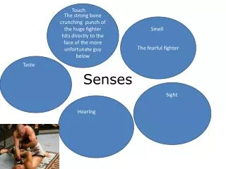
Retina • Made of cells that are light receptors – photoreceptors • Rods and cones • Rods – black and white • Cones - color • Color blindness • Lack of cones
Colorblindness A genetic trait that affects boys more than girls. The location of the gene is on the X chromosome
Image Formation • Seeing is like taking a picture • The object must be focused on a “film” – retina • The correct amount of light must be present – pupil Light bends - refraction
Refraction • When light bends as it moves between two mediums – air and water • Images on the retina are upside-down and have right-to-left reversal
Image Formation Process • Light hits the cornea and is bent • Light leaves the cornea and is bent again • Light enters the lens where it is focused on the retina • So why don’t we see everything upside-down? • Very early on the brain “learns” how to coordinate the images and make them correct
Accommodation • Most of the focusing is done by the cornea • 25% must be done by the lens • Our lens is convex on both sides in order to produce clear images • The lens will increase its curvature in order to focus all images exactly
Animations • http://www.bbc.co.uk/science/humanbody/body/factfiles/sight/sight_animation.shtml • http://www.biologymad.com/resources/eye.swf
Abnormalities and Changes • Presbyopia – lens loses elasticity with age • Around age 40 the lens can’t focus near images and people need glasses • Normal eye – emmetropic – can reflect images perfectly of objects 20 ft away • Myopia - near-sightedness • Eyeball is too long for the focusing power of the lens or lens is thicker than normal • Hyperopia – far-sightedness • Eyeball is too short for the focusing power of the lens or lens is thinner than normal