Download
1 / 25
310 likes | 1.73k Views
Learn about the differences, symptoms, and management of episcleritis and scleritis in ophthalmology. Understand the clinical features, classifications, and associated systemic conditions of these eye diseases.

E N D
Episcleritis and Scleritis Dr Anupam Associate Professor Ophthalmology
Applied Anatomy • Sclera forms the posterior five-sixth opaque part of the of the eyeball. • Its whole outer surface is covered by Tenon's capsule and bulbar conjunctiva. • Its inner surface lies in contact with choroid with a potential suprachoroidal space in between.
It is generally thinner in children & females. • Sclera is thickest posteriorly (1mm) and is thinnest at the insertion of extraocular muscles (0.3 mm). • Lamina cribrosa is a sieve-like sclera from which fibres of optic nerve pass.
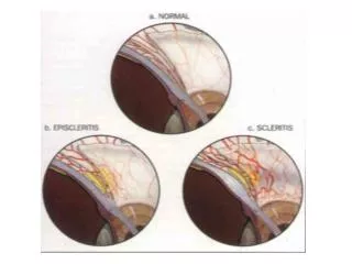
Microscopic structure 1. Episcleral tissue. 2. Sclera proper. 3. Lamina fusca. Vasculature: 1. Conjunctival vessels 2. Episcleral vessels 3. Deep vascular plexus
EPISCLERITIS • Benign recurrent inflammation of the episclera, • Common • Benign • Self-limiting • Recurrent • Never progresses to scleritis • Rarely associated with systemic disease
Etiology • Exact etiology is not known. • It is found in association with gout, rosaceaand psoriasis. • It has also been considered a hypersensitivity reaction to endogenous tubercular or streptococcal toxins. Pathology • Histologically, there occurs localisedlymphocytic infiltration of episcleral tissue associated with oedema and congestion of overlying Tenon's capsule and conjunctiva.
Clinical picture • Symptoms. • Redness, mild ocular discomfort, burning sensation or foreign body sensation. • Rarely, mild photophobia and lacrimation may occur.
Signs • Simple or diffuse episcleritis – Sectoral redness – Diffuse redness – Resolves in 1-2 weeks • Nodular episcleritis – Focal, raised, nodular – Sclera uninvolved – Longer to resolve
Vessels remain radial and mobile • Palpation of the globe often elicits marked tenderness in scleritis, but generally not in episcleritis. • Phenylephrine diagnostic test: Hyperemia usually blanches with topical phenylephrine (2.5%) in episcleritis but not in scleritis.
Management • Mild cases – Usually no specific Rx – If discomfort • Lubricant • Topical NSAID egacular (keterolactrimethamine) • Mild topical corticosteroid Or systemic Ibuproven/aspirin Investigate in recurrent cases.
SCLERITIS • Scleritis refers to a chronic inflammation of the sclera proper. It is a comparatively serious disease which may cause visual impairment and even loss of the eye if treated inadequately. • Relatively rarer than episcleritis • Usually bilateral • More common in females • Associated with connective tissue disorders in upto 50% of cases. • Granulomatous inflammation • Mild to blinding spectrum
Clinical classification of Scleritis It can be classified as follows: I. Anterior scleritis (98%) 1. Non-necrotizing scleritis (85%) (a) Diffuse (b) Nodular 2. Necrotizing scleritis (13%) (a) with inflammation (b) without inflammation (scleromalaciaperforans) II. Posterior scleritis (2%)
Associated systemic conditions • Rheumatoid Arthritis • 1:200 develop scleritis • Connective Tissue Disease • Wegener granulomatosis • Systemic lupus erythematosus • Polyarteritisnodosa • Ankylosingspondilytis
Associated systemic conditions • Herpes Zoster Ophthalmicus • Metabolic disorders like gout and thyrotoxicosis • Granulomatous diseases like • Tuberculosis, • Syphilis, • Sarcoidosis, • Leprosy • Miscellaneous • Surgically induced • Infectious • Idiopathic
Clinical features • Symptoms • Pain • Redness • Photophobia • Lacrimation • Diminution of vision
Clinical features • Signs 1. Non-necrotizing anterior diffuse scleritis. • Commonest variety, • Widespread inflammation involving a quadrant or more of the anterior sclera. • The involved area is raised and salmon pink to purple in colour
Clinical features • Signs 2. Necrotizing anterior nodular scleritis. • characterised by one or two hard, purplish elevated scleral nodules, usually situated near the limbus • Sometimes, the nodules are arranged in a ring around the limbus (annular scleritis).
Clinical features Signs 3. Necrotizing scleritis with inflammation. • The affected necrosed area is thinned out and sclera becomes transparent and ectatic with uveal tissue shining through it. • It is usually associated with anterior uveitis. 4. Anterior necrotizing scleritis without inflammation (scleromalaciaperforans). • Usually associated with seropositive RA • Painless scleral thinning due to ischaemia.
Posterior Scleritis • Defined as primarily arising posterior to the equator • Painful or painless diminution of vision • Proptosis • Restricted ocular movements • Disc or macular edema • Choroidal folds or detachment • Uveal effusion syndrome • Retinal detachment • Scleral thickening seen on CT or USG B scan.
Investigation 1. TLC, DLC and ESR 2. Serum levels of complement (C3), immune complexes, rheumatoid factor, antinuclear antibodies and L.E cells for an immunological survey. 3. FTA - ABS, VDRL for syphilis. 4. Serum uric acid for gout. 5. Urine analysis. 6. Mantoux test. 7. X-rays of chest, paranasal sinuses, sacroiliac joint and orbit to rule out foreign body especially in patients with nodular scleritis.
Management • (A) Non-necrotisingscleritis. • Steroid eye drops and systemic indomethacin 100 mg daily for a day and then 75 mg daily. • B) Necrotisingscleritis. It is treated by topical steroids and heavy doses of oral steroids tapered slowly. • Immuno-suppressive agents like methotrexate or cyclophos-phamide. • Subconjunctival steroids are contraindicated because they may lead to scleral thinning and perforation
STAPHYLOMAS • Ectasia or bulging of the outer coats (cornea, sclera or both) of the eye with incarceration of the uveal tissue. • Due to weakening of the eye wall resulting from any degenerative or inflammatory condition of the same. • Types: • Anterior (involves cornea) • Intercalary (with in 2mm of limbus) • Ciliary (2-8mm behind the limbus • Equatorial (14mm behind the limbus) • Posterior (posterior to equator)
A: Intercalary staphyloma • B. Ciliarystaphyloma • Equatorial staphyloma • Posterior staphyloma
Management • Treat the underlying cause like, scleritis, RA, vit A def or cornal ulcer. • Local excision and patch graft of cornea or sclera • Enucleation with implant.