Download
1 / 63
630 likes | 682 Views
Learn about the early development stages, structure, functions, and pathology of the placenta during pregnancy. Discover how the placenta supports fetal growth and maternal health.

E N D
The placenta Prepared by: Areefa SM Albahri
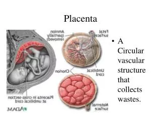
Introduction • The placenta is a complex organ, which originates from the trophoblastic layer of the fertilized ovum. When fully developed it serves as the interface between the mother and the developing fetus carrying out functions that the fetus is unable to perform for itself during intrauterine life. The survival of the fetus depends upon the placenta's integrity and efficiency.
Early development • Pre-implantation Embryonic Development • Following fertilization, the zygote undergoes five or six rapid mitotic cell divisions. Although each cleavage results in more cells. • Each daughter cell produced by cleavage is called a blastomere (blastos = “germ,” in the sense of a seed or sprout). • The inner mass of embryonic cells is totipotent during this stage, meaning that each cell has the potential to differentiate into any cell type in the human body.
Approximately 3 days after fertilization, a 16-cell conceptus reaches the uterus. The cells that had been loosely grouped are now compacted and look more like a solid mass. The name given to this structure is the morula (morula = “little mulberry”). • Once inside the uterus, the conceptus floats freely for several more days. It continues to divide, creating a ball of approximately 100 cells, and consuming nutritive endometrial secretions called uterine milk while the uterine lining thickens. The ball of now tightly bound cells starts to secrete fluid and organize themselves around a fluid-filled cavity, the blastocoel. • At this developmental stage, the conceptus is referred to as a blastocyst. • Within this structure, a group of cells forms into an inner cell mass, which is fated to become the embryo. • The cells that form the outer shell are called trophoblasts (trophe = “to feed” or “to nourish”). • These cells will develop into the chorionic sac and the fetal portion of the placenta (the organ of nutrient, waste, and gas exchange between mother and the developing offspring).
Early development • By 8 days, the trophoblasts begin to make human chorionic gonadotrophin (hCG), a hormone that ensures that the endometrium will be receptive to the implanting embryo. • Implantation • Once the blastocyst makes contact with the endometrium, the trophoblast layer adheres to the endometrial surface and the process of placentation begins. By 10 days, the endometrium layer called the decidua.
Chorionic villi • Initially the blastocyst appears to be covered by fine, downy hair, which consists of the projections from the trophoblastic layer. These proliferate and branch from about 3 weeks after fertilization, forming the chorionic villi. The villi become most profuse in the area where the blood supply is richest, the deciduabasalis.
Chorionic villi • The villi penetrate the decidua, opening them up to form a lake of maternal blood in which they float. The maternal blood circulates slowly, enabling the villi to absorb food and oxygen and excrete waste. These are known as the nutritive villi.
The mature placenta • The placenta is completely formed and functioning 10 weeks after fertilization. Between 12 and 20 weeks' gestation, the placenta weighs more than the fetus. Later in pregnancy, some of the fetal organs, such as the liver, begin to function, so the cytotrophoblast gradually degenerate and this allows easier exchange of oxygen and carbon dioxide.
Placental pathology and maternal disease can affect this ratio: such being diabetes, pre-eclampsia, pregnancy-induced hypertension or intrauterine growth restriction (IUGR)
The weight of the placenta may be affected by physiological or active management of the third stage of labour owing to the varying amounts of fetal blood retained in the vessels. The placenta is no longer routinely weighed in clinical practice; however some maternity units may do so as part of clinical trials and research activities.
The maternal surface of the placenta (i.e. the basal plate) is dark red in colour due to maternal blood and partial separation of the basal decidua • The surface is arranged in up to 40 cotyledons (lobes), which are separated by sulci (furrows), into which the decidua dips down to form septa (walls). The cotyledons are made up oflobules, each of which contains a single villus with its branches.
Functions of Placenta • a variety of functions for the developing fetus. 1. Storage • The placenta metabolizes glucose, stores it in the form of glycogen and reconverts it to glucose as required. It can also store iron and the fat-soluble vitamins.
Endocrine function • The many and varied endocrine functions of the placenta are complex, requiring maternal and fetal input. Both types of trophoblasts produce steroidal hormones (oestrogens and progesterone) in addition to many placental protein hormones necessary for pregnancy
Steroid hormones • There are three important oestrogens: oestrone, oestradiol and oestriol. Both maternal and fetal adrenal production provide precursors for oestrogen production by the placenta. Pregnalone sulphate is converted to oestriol by the feto-placental unit from 6 to 12 weeks onwards, rising steadily until term.
Oestrogens influence uterine blood flow, enhance ribonucleic acid (RNA) and protein synthesis and aid growth of uterine muscle. • They also increase the size and mobility of the maternal nipple and cause alveolar and duct development of the breast tissue.
Progesteroneproduction is maintained by the corpus luteum for approximately 8 weeks until the placenta takes over this function and is dependent on maternal cholesterol stores. • Progesterone is thought to play an important part in immunosuppression to maintain the pregnancy
It maintains the myometrium in a quiescent state, during pregnancy. • It is involved in preparing breast tissue during pregnancy and when levels reduce aher birth of the placenta, prolactin stimulates lactation.
Protein hormones • Human chorionic gonadotrophin (hCG) is • produced under the influence of placental • gonadotrophic releasing hormone (GnRH) by the trophoblasts. • Initially it is present in very large quantities, with peak levels being achieved between the 7th and 10th week, but these gradually reduce as the pregnancy advances. • The function of hCG is to stimulate the corpus luteum to produce mainly progesterone.
It also increases fetal leydig cells to affect male sexual development prior to fetal luteinizing hormone (LH) production (Kay et al 2011). Human chorionic gonadotrophin forms the basis of the many pregnancy tests available, as it is excreted in the mother's urine.
Human placental lactogen (hPL) is sometimes known as human chorionic somatomammotropin hormone (hCS) as it not only stimulates somatic growth but also stimulates proliferation of breast tissue in preparation for lactation.
In early pregnancy, HPL stimulates food intake and weight gain, mobilizing free fafy acids, and functions with prolactin to increase circulating insulin levels • HPL is no longer considered the primary agent of insulin resistance as other growth hormones, such as human placental growth hormone (hPGH), appear to be the main determinants for this. Levels of hPL have been used as a screening tool in pregnancy to assess placental function.
. Respiration • the fetus must obtain oxygen and excrete carbon dioxide through the placenta. Oxygen from the mother's haemoglobin passes into the fetal blood by simple diffusion; similarly, the fetus gives off carbon dioxide into the maternal blood.
2. Nutrition • The fetus needs nutrients for growth and development. For instance, amino acids and , glucose for energy and growth, calcium and phosphorus for bones and teeth, and iron and other minerals for blood formation. These nutrients are actively transferred from the maternal to the fetal blood through the walls of the villi.
Nutrition • Water, vitamins and minerals also pass to the fetus. Fats and fat-soluble vitamins (A, D and E) cross the placenta only with difficulty and mainly in the later stages of pregnancy. Some substances, including amino acids, are found at higher levels in the fetal blood than in the maternal blood.
3. Storage • The placenta metabolizes glucose, stores it in the form of glycogen and reconverts it to glucose as required. It can also store iron and the fat-soluble vitamins.
4. Excretion • The main substance excreted from the fetus is carbon dioxide. Bilirubin will also be excreted as red blood cells are replaced relatively frequently. There is very little tissue breakdown apart from this and the amounts of urea and uric acid excreted are vary
5. Protection • The placenta provides a limited barrier to infection. Few bacteria can penetrate like syphilis and the tubercle bacillus. However, substances including alcohol, some chemicals associated with smoking cigarettes and several types of viruses, such as human cytomegalovirus and rubella are not filtered out. These substances can cross the placental barrier freely and may cause congenital abnormalities.
Although some drugs will cross the placental barrier to the fetus, many will be harmless and others, such as antibiotics administered to a pregnant woman with syphilis, are positively beneficial. • Towards the end of pregnancy, antibodies in the form of immunoglobulin G (IgG) are transferred across the placental barrier to the fetus conferring passive immunity on the baby for the first 3 months of extrauterine life. However, it is important to realize that only those antibodies that the mother herself possesses can be passed on.
6. Endocrine • Oestrogens are growth stimulating hormones, which are secreted throughout pregnancy. They are produced by the placenta as the activity of the corpus luteum declines, the fetus providing the placenta with the vital precursors for their production. The amount of oestrogen produced (measured as urinary or serum oestriol) is an index of fetoplacental well-being.
Human placental lactogen (hPL) has a role in glucose metabolism in pregnancy. As the level of hCG falls, so the level of hPL rises and continues to do so throughout pregnancy. Monitoring the level of hPL with the intention of assessing placental function has been disappointing in predicting fetal outcome
Placental circulation • Maternal blood is discharged into the intervillous space by 80–100 spiral arteries in the deciduabasalis and flows slowly around the villi. There are about 150 mL of maternal blood in the intervillousspaces, which is exchanged 3 or 4 times/min.
Placental circulation • Fetal blood, is pumped by the fetal heart towards the placenta along the umbilical arteries and transported along their branches to the capillaries of the chorionic villi where exchange of nutrients takes place between the mother and fetus.
The membranes • There are two membranes, an outer membrane, the chorion, and an inner membrane, the amnion. As long as the membranes remain intact, they protect the fetus against ascending bacterial infection. • When first formed the amnion is in contact with the embryo, but 4–5 weeks after conception the amniotic fluid begins to accumulate within it.
Amniotic fluid • Amniotic fluid is a clear alkaline and slightly yellowish liquid contained within the amniotic sac. • Functions • Amniotic fluid allowing for the growth and free movement of the fetus and permitting symmetrical musculoskeletal development.
Origin • The source of amniotic fluid both fetal and maternal. • Some fluid is Maternal plasma exuded from maternal vessels in the decidua and some from fetal vessels in the placenta. It is secreted by the amnion, At first mainly consist of water & electrolytes. • by 14wk Protein, lipid, and phospholipid. • Fetal urine also contributes to the volume from the 10th week of gestation onwards. The water in amniotic fluid is exchanged approximately 3-hourly.
Constituents • Amniotic fluid consists of 99% water with the remaining 1% being dissolved solid matter including food substances and waste products. • In addition, the fetus sheds skin cells, vernixcaseosa and lanugo into the fluid. • Abnormal constituents of the liquor, such as meconium in the case of fetal compromise, may give valuable diagnostic information about the condition of the fetus. Aspiration of amniotic fluid for examination is termed amniocentesis.
Volume • During pregnancy, amniotic fluid increases in volume as the fetus grows. The volume is greatest at approximately 38 weeks' gestation, when there is about 1 L. It then diminishes slightly until term, when approximately 800 mL remains. • There are very wide variations in the amount, however if the total amount of fluid exceeds 1500 mL, the condition is known as polyhydramnios and if less than 300 mL, oligohydramnios. Such abnormalities are often associated with congenital malformations of the fetus.
The umbilical cord • The umbilical cord, which extends from the fetal surface of the placenta to the umbilical area of the fetus, is formed by the 5th week of pregnancy. • It originates from the duct that forms between the amniotic sac and the yolk sac and transmits the umbilical blood vessels. • Functions • The umbilical cord transports oxygen and nutrients to the developing fetus, and removes waste products.
Structure • The umbilical cord contains two arteries and one vein, which are continuous with the blood vessels in the chorionic villi of the placenta. The blood vessels are enclosed and protected by Wharton's jelly, a gelatinous substance formed from mesoderm. • The whole cord is covered in a layer of amnion that is continuous with that covering the placenta. There are no nerves in the umbilical cord, so cutting it following the birth of the baby is not painful.
Measurements • The cord is approximately 1–2 cm in diameter and 40–50 cm in length. This length is sufficient to allow for the birth of the baby without applying any traction to the placenta. • A cord is considered short when it measures <40 cm. There is no specific agreed length for describing a cord as too long, but the disadvantages of a very long cord are that it may become wrapped round the neck or body of the fetus or become knotted. Either event could result in occlusion of the blood vessels, especially during labour.
Compromise of the fetal blood flow through the umbilical cord vessels can have serious deleterious effects on the health of the fetus and newborn. True knots should always be noted on examination of the cord, but they must be distinguished from false knots, which are lumps of Wharton's jelly on the side of the cord and are not significant.
The placenta at term • At term, the placenta is a round flat mass about 20 cm in diameter and 2.5 cm thick at its centre. • It weighs approximately(470g) one-sixth of the baby's weight, although this proportion may be affected by the time at which the cord is clamped owing to the varying amounts of fetal blood retained in the vessels.