Osteochondritis Dissicans
120 likes | 775 Views
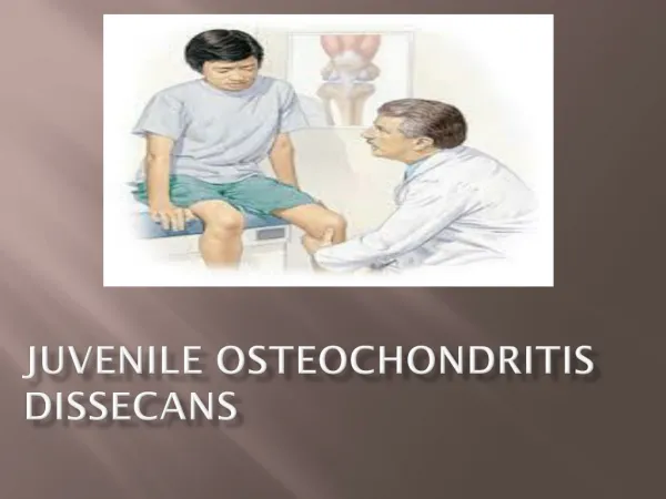
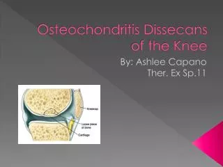
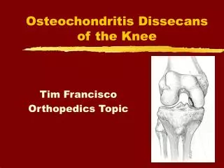
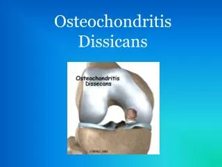
Osteochondritis Dissicans. What is it?. Disorder of an ossification site Can affect one or more joints Affects the: Subchondral layer Articular cartilage Bone fragment and articular cartilage separate from underlying bone Fragments create loose bodies in the joint. Classification.

Osteochondritis Dissicans
E N D
Presentation Transcript
What is it? • Disorder of an ossification site • Can affect one or more joints • Affects the: • Subchondral layer • Articular cartilage • Bone fragment and articular cartilage separate from underlying bone • Fragments create loose bodies in the joint
Classification • Based on bone maturity • Juvenile lesions • Open distal femoral physes • Adolescents • Closing distal femoral physes • Adults • Fully closed distal femoral physes • Also based on radiographic imaging • Size and location of the lesion • Stability and loose fragments
Etiology • Genetics-hereditary • Tibia vara • Legg-Calve-Perthes disease • Stickler’s syndrome • Epiphyseal dysplasia • Vascular • Trauma (40%) • Repetitive/persistent microtrauma
Commonly affected areas • Medial femoral condyle • Lateral portion • Inferiocentral • Lateral femoral condyle • Anterior • Inferiocentral • Occurs bilaterally in many cases
Population at risk • 10-15 years old • Males > females
Symptoms • Pain with activity • Swelling • Giving way • Wilson’s sign • Pain increased with passive knee extension and internal tibial rotation • Pain decreases with tibial external rotation
Diagnosis • Confirmed by radiographic images
Management • Depends on the extent of the damage and age • Non-operative management • Activity modification • Protected weight bearing • Immobilization 4-6 weeks followed by • Quadriceps strengthening • Gradual return to activity • Surgical • If loose bodies are present or unsuccessful conservative treatment
References • Goodman, Catherine C., Fuller, Kenda. 2009. Pathology: Implications for the Physical Therapist, 3rd ed. Saunders/Elsevier. St. Louis, Mo. • DeLee, Jesse C., Drez, David Jr. 2003. DeLee & Drez’sOrthopaedic Sports Medicine: Principles and Practice, 3rd ed. Saunders. Philadelphia, PA.