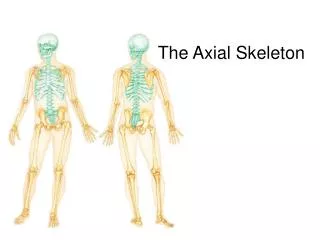
The Axial Skeleton
550 likes | 1.04k Views
The Axial Skeleton. This structure is composed of 27 bones and is formed from cranial and facial bones. The cranial bones protect the brain and allow attachment for the neck and head muscles. The Skull. The facial bone have several functions: Form the frame work for the face

The Axial Skeleton
E N D
Presentation Transcript
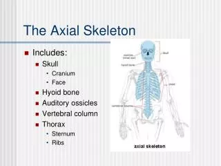
This structure is composed of 27 bones and is formed from cranial and facial bones. The cranial bones protect the brain and allow attachment for the neck and head muscles. The Skull
The facial bone have several functions: Form the frame work for the face Contain cavities for the senses Provided openings for air and food Secure the teeth Anchor the facial muscles The Skull
With the exception of the mandible all bones of the adult skull are interlocked by joints called sutures. The major sutures are the Coronal Sagittal Squamous Lambdoid Sutures
Figure 7.2a The skull: Cranial and facial divisions and fossae. Bones of cranium (cranial vault) Coronal suture Squamous suture Facial bones Lambdoid suture (a) Cranial and facial divisions of the skull
The floor is divided into the anterior , middle and posterior fossae The Floor
Figure 7.2b The skull: Cranial and facial divisons and fossae. Anterior cranial fossa Middle cranial fossa Posterior cranial fossa (b) Superior view of the cranial fossae
Anterior and Posterior Aspects of the Skull • Supraorbital margin is a throw back to our simian cousins • For us it supports our eye brows • The Supraorbital foramen is the path for the supraorbital nerve and vessels
Anterior and Posterior Aspects The superior nuchal and inferior nuchal lines serve as attachment points for muscles and ligaments.
Figure 7.5a Bones of the lateral aspect of the skull, external and internal views. Frontal bone Coronal suture Sphenoid bone (greater wing) Parietal bone Ethmoid bone Temporal bone Lacrimal bone Lacrimal fossa Lambdoid suture Squamous suture Nasal bone Occipital bone Zygomatic bone Zygomatic process Maxilla Occipitomastoid suture External acoustic meatus Alveolar margins Mastoid process Styloid process Mandibular condyle Mandible Mandibular notch Mental foramen Mandibular ramus Coronoid process Mandibular angle (a) External anatomy of the right side of the skull
Figure 7.7b The floor of the cranial cavity. Crista galli Frontal bone Ethmoid bone Cribriform plate Olfactory foramina Anterior cranial fossa Optic canal Foramen rotundum Lesser wing Sphenoid Foramen ovale Greater wing Foramen spinosum Middle cranial fossa Foramen lacerum Hypophyseal fossa of sella turcica Jugular foramen Temporal bone (petrous part) Foramen magnum Posterior cranial fossa View Parietal bone Occipital bone (b) Superior view of the skull, calvaria removed
Figure 7.6b Inferior aspect of the skull, mandible removed. Hard palate Zygomatic arch Foramen ovale Foramen spinosum Foramen lacerum Mandibular fossa Carotid canal Styloid process Mastoid process Jugular foramen Occipital condyle Foramen magnum Superior nuchal line (b) Photo of inferior view of the skull
Figure 7.11a Detailed anatomy of the mandible and the maxilla. Mandibular fossa of temporal bone Temporomandibular joint Mandibular notch Coronoid process Mandibular condyle Mandibular foramen Alveolar margin Ramus of mandible Mental foramen Mandibular angle Body of mandible (a) Mandible, right lateral view
The Hyoid Bone This is a “U” shaped bone. It is not connected to the skull. It forms the base for the tongue.
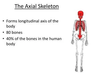
The Spinal Column The vertebral column consists of 26 irregular bones. It provides the main axial support for the skeleton.
Figure 7.16 The vertebral column. C1 Cervical curvature (concave) 7 vertebrae, C1–C7 Spinous process Transverse processes Thoracic curvature (convex) 12 vertebrae, T1–T12 Intervertebral discs Intervertebral foramen Lumbar curvature (concave) 5 vertebrae, L1–L5 Sacral curvature (convex) 5 fused vertebrae sacrum Coccyx 4 fused vertebrae Anterior view Right lateral view
The Spinal Column There are 7 cervical vertebrae 12 Thoracic vertebrae 5 Lumbar vertebrae 5 Sacral vertebrae
Anterior and Posterior Aspects of the Skull You have breakfast at 7
Anterior and Posterior Aspects of the Skull You have lunch at 12
Anterior and Posterior Aspects of the Skull You have dinner at 5
Anterior and Posterior Aspects of the Skull You have to go to the bathroom at 5 am
The Spinal Column The major supporting ligaments are the anterior and posterior longitudinal ligaments.
Figure 7.17a Ligaments and fibrocartilage discs uniting the vertebrae. Intervertebral disc Supraspinous ligament Anterior longitudinal ligament Transverse process Sectioned spinous process Intervertebral foramen Posterior longitudinal ligament Ligamentum flavum Interspinous ligament Anulus fibrosus Nucleus pulposus Inferior articular process Sectioned body of vertebra Median section of three vertebrae, illustrating the composition of the discs and the ligaments
The Spinal Column The anterior ligament attaches to the vertebrae and discs. It prevents hyperextension (bending backward) The posterior ligament is weak and resists hyperflexation.
The Spinal Column The Intervertebral discs accounts for 25% of your height and acts as a shock absorber. A herniated or slip discs is a common cause of back injuries.
Figure 7.17c Ligaments and fibrocartilage discs uniting the vertebrae. Vertebral spinous process (posterior aspect of vertebra) Spinal cord Spinal nerve root Transverse process Herniated portion of disc Anulus fibrosus of disc Nucleus pulposus of disc (c) Superior view of a herniated intervertebral disc
Figure 7.17d Ligaments and fibrocartilage discs uniting the vertebrae. Nucleus pulposus of intact disc Herniatednucleus pulposus (d) MRI of lumbar region of vertebral columnin sagittal section showing herniated disc
The Cervical Vertebrae These are the smallest with C1 and C2 modified for the skull. In general cervical vertebrae have • An oval body • A short spinous process which is split except for C7 • A transverse foramen for the vertebral arteries.
The Thoracic Vertebrae There are12 (T1-T12) These have : 1) Circular vertebral foramen 2)A long spinous process that points downward. 3) Transverse processes have facets for the ribs
Figure 7.20b Posterolateral views of articulated vertebrae. Superior articular process Transverse process Transverse costal facet (for tubercle of rib) Intervertebral disc Body Inferior costal facet (for head of rib) Spinous process Inferior articular process (b) Thoracic vertebrae
The Lumbar Vertebrae There are 5(L1-L5) These have : 1) Spinous process is short & flat 2) Vertebral foramen is triangular 3) articular processes face medially or laterally
Figure 7.20c Posterolateral views of articulated vertebrae. Superior articular process Body Transverse process Intervertebral disc Inferior articular process Spinous process (c) Lumbar vertebrae
The Sacral Vertebrae There are 5 (S1-S5) These are fused and articulates with L5 and the ileum
Figure 7.21 The sacrum and coccyx. Facet of superior articular process Body Sacral canal Ala Sacral promontory Auricular surface Ala Body of first sacral vertebra Median sacral crest Lateral sacral crest Posterior sacral foramina Transverse ridges (sites of vertebral fusion) Sacral hiatus Anterior sacral foramina Coccyx Apex (b) Posterior view Coccyx (a) Anterior view
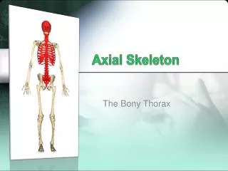
The Thoracic Cage This is composed of the ribs, thoracic vertebrae dorsally and sternum ventrally. Ribs 1-7 are true ribs because they attach directly to the sternum.
The Thoracic Cage This is composed of the ribs, thoracic vertebrae dorsally and sternum ventrally. Ribs 1-7 are true ribs because they attach directly to the sternum. Ribs 8-10 are false ribs because they attach indirectly
The Thoracic Cage This is composed of the ribs, thoracic vertebrae dorsally and sternum ventrally. Ribs 1-7 are true ribs because they attach directly to the sternum. Ribs 8-10 are false ribs because they attach indirectly Ribs 11 & 12 are floating and are NOT attached to the sternum
Figure 7.22 The thoracic cage. Jugular notch Clavicular notch Manubrium Sternal angle Body Sternum Xiphisternal joint Xiphoid process True ribs (1–7) Intercostal spaces Jugular notch Sternal angle False ribs (8–12) Heart Costal cartilage Xiphisternal joint L1 Vertebra Floating ribs (11, 12) Costal margin (b) Midsagittal section through the thorax, showing the relationship of surface anatomical landmarks of the thorax to the vertebral column (a) Skeleton of the thoracic cage, anterior view
Figure 7.23c Ribs. Facets for articulation with vertebrae Articular facet on tubercle Shaft Head Neck Costal angle Junction with costal cartilage Costal groove (c) A typical rib (rib 6, right), posterior view