Download
1 / 20
1.39k likes | 12.99k Views
CERVICAL LYMPHADENOPATHY. Prof. SHANMUGASUNDARAM. P.N Professor and Head Department of General Surgery Kilpauk medical college. Introduction. Enlarged lymph nodes is one of the common presentation in head and neck patients.

E N D
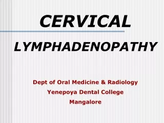
CERVICAL LYMPHADENOPATHY Prof. SHANMUGASUNDARAM. P.N Professor and Head Department of General Surgery Kilpauk medical college
Introduction • Enlarged lymph nodes is one of the common presentation in head and neck patients. • Though they are easily recognizable, considerable clinical skill and expertise is required to categorize these swellings and manage them appropriately. • Recent advances in imaging have considerably improved the diagnostic and therapuetic facility for these patients.
definition • Cervical lymphadenitis is defined as enlarged, inflamed, and tender lymph node(s) of the neck. • Cervical lymphadenopathy refers simply to enlarged lymph node(s) of the neck. However, the two terms often are used interchangeably. • Generalized lymphadenopathy is the enlargement of more than two noncontiguous lymph node regions (ie, cervical and axillary) and is the result of systemic disease.
Clinical claSSIFICATION Cervical lymphadenopathy • Ref: 18th edition of Harrison's Principles of Internal Medicine Table 59–1 malignant Benign Infectious diseases Hematologic: Lymphoma, Hairy cell leukemia Immunological : RA, SLE , Serum sickness Endocrine diseases—hyperthyroidism Lipid storage diseases—Gaucher's, Niemann-Pick, Fabry, Tangier Metastatic Drug hypersensitivity Other causes : Kikuchi's disease (Histiocytic necrotizing lymphadenitis), Rosai-Dorfman disease(Sinus histiocytosis), Kawasaki's disease (Mucocutaneous lymph node syndrome)
Classification of nodes • The neck comprises of more than 300 lymph nodes • They can be classified as the superficial group and the deep group.
Classification of nodes • Superficial group of nodes • Preauricular • Postauricular • Occipital • Preparotid • Facial Adapted from Gray’s textbook of anatomy
Classification of nodes • The deep cervical nodes are classified in terms of important clinical/surgical landmarks. • They are classified into six neck levels. • Each level denotes a specific drainage areas which give important diagnostic clues.
Classification of nodes • Classification by the Memorial of Sloan Kettering. • Primarily used to describe the metastatic deposits from squamous cell carcinoma of head and neck.
SALIENT FEATURES IN HISTORY TAKING • Symptoms such as sore throat, cough, fever, night sweats, fatigue, weight loss, or pain in the nodes should be sought. • The patient's age, sex, occupation, exposure to pets, sexual behavior, a history of tobacco use and use of drugs such as di-phenyl hydantoin are other important historic points • After age 50, the incidence of malignant disorders increases and that of benign disorders decreases.
CLINICAL Examination • Size • Nodes greater than 3 cm are associated with malignancy in adult patients, • Nodes greater than 2 cm in children in the absence of ENT infection and absence of abnormal chest radiograph point to a underlying granulomatous disorder. • Pain/Tenderness • an inflammatory process or suppuration • hemorrhage into the necrotic center of a malignant node
Clinical examination • Consistency • Stony-hard nodes are typically a sign of cancer, usually metastatic. • Very firm, rubbery nodes suggest lymphoma. • Softer nodes are the result of infections or inflammatory conditions. • Suppurant nodes may be fluctuant. • The term “shotty” refers to small nodes that feel like buckshot under the skin, as found in the cervical nodes of children with viral illnesses. • Matting • A group of nodes that feels connected and seems to move as a unit is said to be “matted.” • Nodes that are matted can be either benign (e.g., tuberculosis, sarcoidosis ) or malignant (e.g., metastatic carcinoma or lymphomas).
CLINICAL EXAMINATION • POSTERIOR CERVICAL LYMPH NODE SUPPURATION WITH ABSENCE OF INFLAMATORY SIGNS USUALLY POINT TO TUBERCULOSIS • EXAMINATION OF THYROID FOR PALPABLE LESIONS • VIRCHOW’S NODE • LATERAL ABBERANT THYROID
Clinical examination • Signs usually indicating a systemic disease (LYMPHOMA,STORAGE DISORDERS) • PRESENCE OF SPLENOMEGALY • PRESENCE OF enlarged or ulcerated tonsils. • Thorough ENT examination • to rule out head and neck focus of infection or malignancy.
Investigations to aid diagnosis • ROUTINE LABORATORY INVESTIGATIONS • COMPLETE HEMOGRAM AND PERIPHERAL SMEAR CANNOT BE OVEREMPHASIZED TO DIAGNOSE CLINICAL CONDITIONS LIKE MONONUCLEOSIS OR HEMATOLOGICAL MALIGNANCIES. • IMAGING • NECK ULTRASOUND • CONTRAST ENHANCED COMPUTED TOMOGRAM • TISSUE DIAGNOSIS • FINE NEEDLE ASPIRATION CYTOLOGY. • LYMPH NODE BIOPSY
WHEN TO obtain a tissue biopsy? • ITS A CLINICAL DECISION MADE BY THE TREATING SURGEON. • CLINICALLY MALIGNANT LYMPH NODE. • SUSPECTED TUBERCULOSIS. • A PERSISTENT LYMPHADENOPATHY in the absence of ENT infection or abnormal chest radiograph and NOT RESPONDING TO CONSERVASTIVE MANAGEMENT. • Presence of gross splenomegaly
FNAC versus BIOPSY ?? • OP PROCEDURE • NO NEED FOR ANAESTHESIA • HIGH SENSITIVITY AND SPECIFICITY • LOW COST • CAN BE REPEATED IN THE SAME NODE. • DISADVANTAGE • HISTOLOGICAL EVALUATION IS LIMITED. • SUBTYPING OF LYMPHOMAS IS CURRENTLY DIFFICULT. • GOLD STANDARD TESTING • CAN BE DONE UNDER LOCAL ANAESTHESIA • INDICATED FOR IMMUNOHISTOCHEMISTRY AND SUBTYPING OF LYMPHOMAS. FNAC NODE BIOPSY
Various common clinical conditions • 10 YEAR OLD CHILD PRESENTING WITH UNILATERAL TONSILLAR ENLARGEMENT AND LYUMPHADENOPATHY USUALLY POINT TO LYMPHOMA.
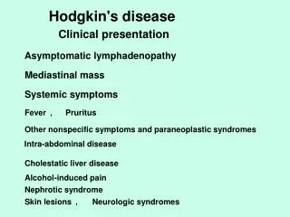
HODGKIN’S LYMPHOMA • YOUNG MALE • PRESENTING WITH NIGHT SWEATS , FEVER, ITCHING { B symptoms} • bilateral neck nodes. • Unilateral Tonsillarenlargemnt
Infectious mononucleosis • Young female • Presenting with fever and throat pain • tonsillar membrane on examination . • Monospot test positive for EBV